კონიუნქტივის მელანოციტური დაზიანების ჰეტეროგენულობის, ფენოტიპური თავისებურებისა და პროგრესიის რისკის მახასიათებლები

ჩამოტვირთვები


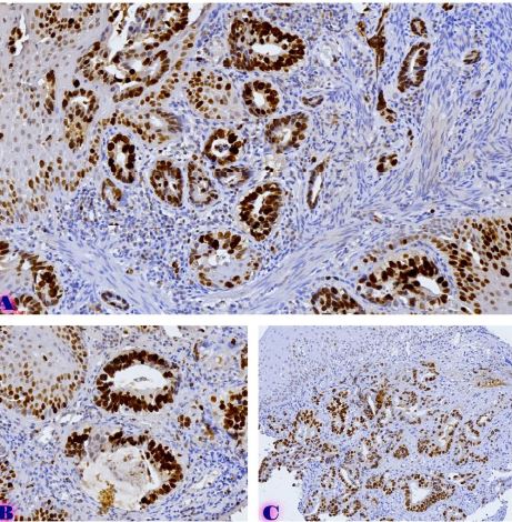
კონიუნქტივის მელანოციტური დაზიანებები წარმოადგენს ჰეტეროგენულ ჯგუფს, რომელიც მოიცავს კეთილთვისებიან, პრეავთვისებიან და ავთვისებიან პროცესებს, მათ შორის ნევუსებს, პირველად შეძენილ მელანოზს (PAM) და კონიუნქტივურ მელანომას. აღნიშნული დაზიანებები განსხვავდება კლინიკური მიმდინარეობით, ჰისტოლოგიური სტრუქტურით, მოლეკულური პროფილითა და ავთვისებიანი ტრანსფორმაციის პოტენციალით. თანამედროვე ოფთალმოპათოლოგიაში განსაკუთრებული მნიშვნელობა ენიჭება იმ ფენოტიპური და მოლეკულური მახასიათებლების იდენტიფიცირებას, რომლებიც ასოცირებულია პროგრესიის, რეციდივისა და მეტასტაზირების რისკთან.
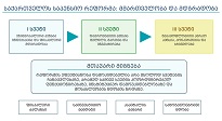
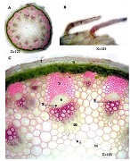
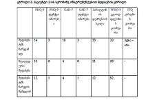
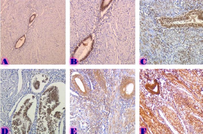
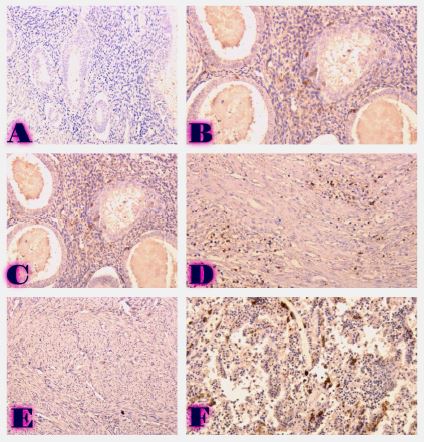
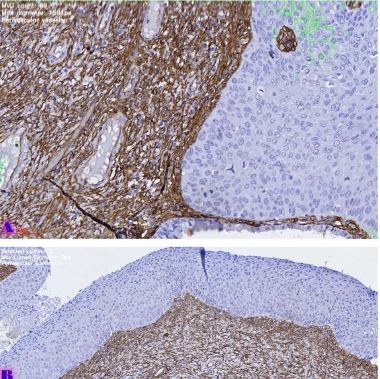
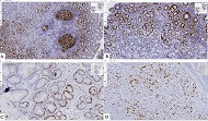
წინამდებარე კრიტიკული მიმოხილვის მიზანია კონიუნქტივის მელანოციტური დაზიანებების თანამედროვე კლასიფიკაციის, ჰისტოლოგიური და იმუნოჰისტოქიმიური თავისებურებების, ეპიდემიოლოგიური მონაცემებისა და პროგრესიის მექანიზმების ანალიზი. განხილულია კონიუნქტივის ნორმალური ჰისტოლოგიური სტრუქტურა, მელანოციტების ბიოლოგიური ფუნქცია, მათი განაწილების თავისებურებები და ისეთი მარკერების მნიშვნელობა, როგორიცაა S-100, HMB-45, Melan-A, Ki-67 და p53. განსაკუთრებული ყურადღება ეთმობა მოლეკულურ ცვლილებებს, მათ შორის BRAF, NRAS და KIT გენების მუტაციებს, რომლებიც დაკავშირებულია მელანოციტური დაზიანებების ავთვისებიან პროგრესიასთან.
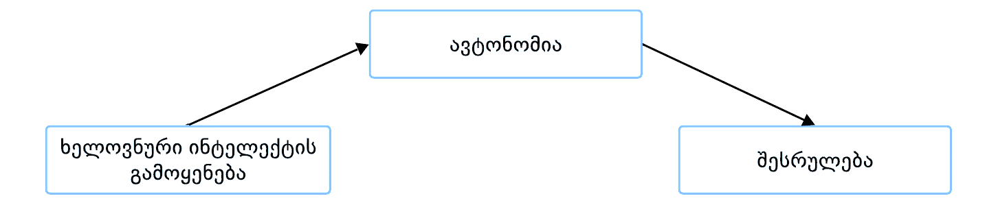
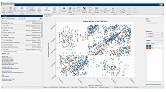
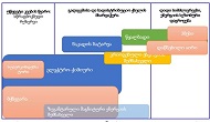
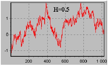
მიმოხილვაში ასევე განხილულია ხელოვნური ინტელექტისა და ციფრული პათოლოგიის როლი დიაგნოსტიკაში, პროგნოზირებასა და რისკის სტრატიფიკაციაში. ხაზგასმულია სიმსივნური მიკროგარემოს, სიმსივნის ინფილტრაციული ლიმფოციტების (TILs), ანგიოგენეზისა და იმუნური რეგულაციის მნიშვნელობა კონიუნქტივური მელანომის ბიოლოგიურ ქცევაში. კონიუნქტივის მელანოციტური დაზიანებების მრავალფეროვნება და იშვიათობა დიაგნოსტიკურ და თერაპიულ სირთულეებს ქმნის, რაც საჭიროებს ინტერდისციპლინურ მიდგომასა და თანამედროვე მოლეკულური მეთოდების ინტეგრაციას. აღნიშნული მიმართულებით შემდგომი კვლევები მნიშვნელოვანია ადრეული დიაგნოსტიკის, ინდივიდუალიზებული მკურნალობისა და პროგნოზის გაუმჯობესებისთვის.
Downloads
M. C. Herwig-Carl, K. U. Loeffler, and H. E. Grossniklaus, “Melanocytoma of the conjunctiva: Clinicopathologic features of three cases,” Ocul. Oncol. Pathol., vol. 5, no. 4, pp. 290–297, Jun. 2019, doi: 10.1159/000496557.
S. Nahon-Estève et al., “Small but challenging conjunctival melanoma: New insights, paradigms and future perspectives,” Cancers (Basel)., vol. 13, no. 22, Nov. 2021, doi: 10.3390/CANCERS13225691
M. Sugiura, K. A. Colby, M. C. Mihm, and A. Zembowicz, “Low-risk and high-risk histologic features in conjunctival primary acquired melanosis with atypia: Clinicopathologic analysis of 29 cases,” American Journal of Surgical Pathology, vol. 31, no. 2, pp. 185–192, Feb. 2007, doi: 10.1097/01.PAS.0000213339.32734.64.
“Melanocytic lesions of the conjunctiva: an up-to-date review - ScienceDirect.” Accessed: May 28, 2026. [Online]. Available: https://www.sciencedirect.com/science/article/abs/pii/S1756231723001652
C. L. Shields, S. Kaliki, S. A. Al-Dahmash, S. E. Lally, and J. A. Shields, “American joint committee on cancer (AJCC) clinical classification predicts conjunctival melanoma outcomes,” Ophthalmic Plast. Reconstr. Surg., vol. 28, no. 5, pp. 313–323, Sep. 2012, doi: 10.1097/IOP.0B013E3182611670.
R. Folberg, I. W. McLean, and L. E. Zimmerman, “Primary acquired melanosis of the conjunctiva,” Hum. Pathol., vol. 16, no. 2, pp. 129–135, 1985, doi: 10.1016/S0046-8177(85)80061-7.
S. Seregard, “Pigmented spindle cell naevus of Reed presenting in the conjunctiva,” Acta Ophthalmol. Scand., vol. 78, no. 1, pp. 104–106, Feb. 2000, doi: 10.1034/J.1600-0420.2000.078001104.X.
C. L. Shields, H. Demirci, J. A. Shields, and C. Spanich, “Dramatic regression of conjunctival and corneal acquired melanosis with topical mitomycin C [2],” British Journal of Ophthalmology, vol. 86, no. 2, pp. 244–245, 2002, doi: 10.1136/BJO.86.2.244.
T. Vandenboom et al., “Genomic fusions in pigmented spindle cell nevus of reed,” American Journal of Surgical Pathology, vol. 42, no. 8, pp. 1042–1051, Aug. 2018, doi: 10.1097/PAS.0000000000001074.
A. Díaz et al., “Pigmented spindle cell nevus: Clues for differentiating it from spindle cell malignant melanoma. a comprehensive survey including clinicopathologic, immunohistochemical, and FISH studies,” American Journal of Surgical Pathology, vol. 35, no. 11, pp. 1733–1742, Nov. 2011, doi: 10.1097/PAS.0B013E318229CF66.
J. H. Francis, H. E. Grossniklaus, L. A. Habib, B. Marr, D. H. Abramson, and K. J. Busam, “BRAF, NRAS, and GNAQ mutations in conjunctival melanocytic nevi,” Invest. Ophthalmol. Vis. Sci., vol. 59, no. 1, pp. 117–121, Jan. 2018, doi: 10.1167/IOVS.17-22517.
R. Folberg and C. I. W. McLean, “Primary acquired melanosis and melanoma of the conjunctiva: Terminology, classification, and biologic behavior,” Hum. Pathol., vol. 17, no. 7, pp. 652–654, 1986, doi: 10.1016/S0046-8177(86)80175-7.
R. Folberg, “Naming the Precursors of Conjunctival Melanoma,” Am. J. Ophthalmol., vol. 162, pp. 1–2, Feb. 2016, doi: 10.1016/J.AJO.2015.11.037.
T. Milman et al., “Validation of the Newly Proposed World Health Organization Classification System for Conjunctival Melanocytic Intraepithelial Lesions: A Comparison with the C-MIN and PAM Classification Schemes,” Am. J. Ophthalmol., vol. 223, pp. 60–74, Mar. 2021, doi: 10.1016/J.AJO.2020.10.020.
G. S. Negretti, K. A. Roelofs, B. Damato, M. Sagoo, S. Parvizi, and V. M. L. Cohen, “The natural history of conjunctival naevi in children and adolescents,” Eye (Basingstoke), vol. 35, no. 9, pp. 2579–2584, Sep. 2021, doi: 10.1038/S41433-020-01273-4.
H. S. Mudhar et al., “PRAME expression by immunohistochemistry and reverse transcription quantitative PCR in conjunctival melanocytic lesions—a comprehensive clinicopathologic study of 202 cases and correlation of cytogenetics with PRAME expression in challenging conjunctival…,” Hum. Pathol., vol. 134, pp. 1–18, Apr. 2023, doi: 10.1016/J.HUMPATH.2023.02.002.
G. Virgili et al., “Incidence and Survival of Patients with Conjunctival Melanoma in Europe,” JAMA Ophthalmol., vol. 138, no. 6, pp. 601–608, Jun. 2020, doi: 10.1001/JAMAOPHTHALMOL.2020.0531.
T. A. Weppelmann, K. T. Zimmerman, and V. Rashidi, “Trends in Incidence of Conjunctival Melanoma in the US,” JAMA Netw. Open, vol. 5, no. 10, Oct. 2022, doi: 10.1001/JAMANETWORKOPEN.2022.37229.
T. J. LIESEGANG, “Pigmented Conjunctival and Scleral Lesions,” Mayo Clin. Proc., vol. 69, no. 2, pp. 151–161, 1994, doi: 10.1016/S0025-6196(12)61042-8.
I. Sayed-Ahmed et al., “Blue Nevi of the Ocular Surface: Clinical Characteristics, Pathologic Features, and Clinical Course,” Ophthalmology, vol. 125, no. 8, pp. 1189–1198, Aug. 2018, doi: 10.1016/J.OPHTHA.2018.02.006.
L. Levecq, P. De Potter, and J. Jamart, “Conjunctival Nevi. Clinical Features and Therapeutic Outcomes,” Ophthalmology, vol. 117, no. 1, pp. 35–40, Jan. 2010, doi: 10.1016/J.OPHTHA.2009.06.018.
D. Šekoranja, G. Hawlina, and J. Pižem, “PRAME expression in melanocytic lesions of the conjunctiva,” Histopathology, vol. 79, no. 6, pp. 989–996, Dec. 2021, doi: 10.1111/HIS.14452.
C. L. Shields, A. Fasiudden, A. Mashayekhi, and J. A. Shields, “Conjunctival Nevi: Clinical Features and Natural Course in 410 Consecutive Patients,” Archives of Ophthalmology, vol. 122, no. 2, pp. 167–175, Feb. 2004, doi: 10.1001/ARCHOPHT.122.2.167.
F. A. Jakobiec, P. Bhat, and K. A. Colby, “Immunohistochemical studies of conjunctival nevi and melanomas,” Archives of Ophthalmology, vol. 128, no. 2, pp. 174–183, Feb. 2010, doi: 10.1001/ARCHOPHTHALMOL.2009.394.
F. A. Jakobiec, K. Colby, A. M. Bajart, S. J. Saragas, and A. Moulin, “Immunohistochemical studies of atypical conjunctival melanocytic nevi,” Archives of Ophthalmology, vol. 127, no. 8, pp. 970–980, Aug. 2009, doi: 10.1001/ARCHOPHTHALMOL.2009.171.
A. Zembowicz, R. V. Mandal, and P. Choopong, “Melanocytic lesions of the conjunctiva,” Arch. Pathol. Lab. Med., vol. 134, no. 12, pp. 1785–1792, Dec. 2010, doi: 10.5858/2009-0522-RAR.1.
Y. Y. Huang, S. M. Hrycaj, M. P. Chan, A. M. Stagner, R. M. Patel, and S. C. Bresler, “PRAME Expression in Junctional Melanocytic Proliferations of the Conjunctiva: A Potential Biomarker for Primary Acquired Melanosis/Conjunctival Melanocytic Intraepithelial Lesions,” American Journal of Dermatopathology, vol. 44, no. 10, pp. 734–740, Oct. 2022, doi: 10.1097/DAD.0000000000002201.
E. Zamir, H. Mechoulam, A. Micera, F. Levi-Schaffer, and J. Pe’er, “Inflamed juvenile conjunctival naevus: Clinicopathological characterisation,” British Journal of Ophthalmology, vol. 86, no. 1, pp. 28–30, 2002, doi: 10.1136/BJO.86.1.28.
K. Svedberg, “Recurrence of Primary Acquired Melanosis and Conjunctival Intraepithelial Neoplasia,” Ocul. Oncol. Pathol., vol. 8, no. 4–6, pp. 236–241, Feb. 2023, doi: 10.1159/000526985.
R. Folberg, I. W. McLean, and L. E. Zimmerman, “Malignant melanoma of the conjunctiva,” Hum. Pathol., vol. 16, no. 2, pp. 136–143, 1985, doi: 10.1016/S0046-8177(85)80062-9.
J. M. McDonnell, Y. Y. Sun, and D. Wagner, “HMB-45 Immunohistochemical Staining of Conjunctival Melanocytic Lesions,” Ophthalmology, vol. 98, no. 4, pp. 453–458, 1991, doi: 10.1016/S0161-6420(91)32281-4.
J. A. Van Ipenburg, J. Damman, D. Paridaens, and R. M. Verdijk, “Histopathological and Molecular Features of a Conjunctival Caruncular Deep Penetrating Nevus,” Ocul. Oncol. Pathol., vol. 6, no. 4, pp. 293–296, Aug. 2020, doi: 10.1159/000504966.
საავტორო უფლებები (c) 2026 ქართველი მეცნიერები

ეს ნამუშევარი ლიცენზირებულია Creative Commons Attribution-NonCommercial-NoDerivatives 4.0 საერთაშორისო ლიცენზიით .