Heterogeneity, Phenotypic Characteristics, and Risk Features of Malignant Progression in Conjunctival Melanocytic Lesions

Downloads




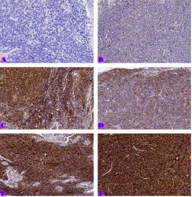
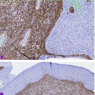
Conjunctival melanocytic lesions represent a heterogeneous group of benign, premalignant, and malignant entities, including conjunctival nevi, primary acquired melanosis (PAM), and conjunctival melanoma. These lesions demonstrate considerable variability in clinical behaviour, histopathological architecture, molecular profile, and malignant transformation potential. In modern ophthalmic pathology, increasing attention is directed toward identifying phenotypic and molecular indicators associated with progression, recurrence, and metastatic risk. This critical review aims to analyse the current classification, histological and immunohistochemical characteristics, epidemiological aspects, and mechanisms of progression of conjunctival melanocytic lesions. The review discusses the normal histological structure of the conjunctiva, the biological role and distribution of melanocytes, and the diagnostic significance of markers such as S-100, HMB-45, Melan-A, Ki-67, and p53. Particular emphasis is placed on molecular alterations, including BRAF, NRAS, and KIT mutations, which are associated with malignant progression.
Additionally, the review highlights the emerging role of artificial intelligence and digital pathology in diagnostic assessment, prognostic evaluation, and risk stratification. The importance of the tumor microenvironment, tumour-infiltrating lymphocytes (TILs), angiogenesis, and immune regulation in the biological behaviour of conjunctival melanoma is also discussed. The heterogeneity and rarity of conjunctival melanocytic lesions continue to create significant diagnostic and therapeutic challenges, necessitating interdisciplinary approaches and integration of modern molecular technologies. Further research in this field is essential for improving early diagnosis, individualised treatment strategies, and patient prognosis.
Downloads
M. C. Herwig-Carl, K. U. Loeffler, and H. E. Grossniklaus, “Melanocytoma of the conjunctiva: Clinicopathologic features of three cases,” Ocul. Oncol. Pathol., vol. 5, no. 4, pp. 290–297, Jun. 2019, doi: 10.1159/000496557.
S. Nahon-Estève et al., “Small but challenging conjunctival melanoma: New insights, paradigms and future perspectives,” Cancers (Basel)., vol. 13, no. 22, Nov. 2021, doi: 10.3390/CANCERS13225691
M. Sugiura, K. A. Colby, M. C. Mihm, and A. Zembowicz, “Low-risk and high-risk histologic features in conjunctival primary acquired melanosis with atypia: Clinicopathologic analysis of 29 cases,” American Journal of Surgical Pathology, vol. 31, no. 2, pp. 185–192, Feb. 2007, doi: 10.1097/01.PAS.0000213339.32734.64.
“Melanocytic lesions of the conjunctiva: an up-to-date review - ScienceDirect.” Accessed: May 28, 2026. [Online]. Available: https://www.sciencedirect.com/science/article/abs/pii/S1756231723001652
C. L. Shields, S. Kaliki, S. A. Al-Dahmash, S. E. Lally, and J. A. Shields, “American joint committee on cancer (AJCC) clinical classification predicts conjunctival melanoma outcomes,” Ophthalmic Plast. Reconstr. Surg., vol. 28, no. 5, pp. 313–323, Sep. 2012, doi: 10.1097/IOP.0B013E3182611670.
R. Folberg, I. W. McLean, and L. E. Zimmerman, “Primary acquired melanosis of the conjunctiva,” Hum. Pathol., vol. 16, no. 2, pp. 129–135, 1985, doi: 10.1016/S0046-8177(85)80061-7.
S. Seregard, “Pigmented spindle cell naevus of Reed presenting in the conjunctiva,” Acta Ophthalmol. Scand., vol. 78, no. 1, pp. 104–106, Feb. 2000, doi: 10.1034/J.1600-0420.2000.078001104.X.
C. L. Shields, H. Demirci, J. A. Shields, and C. Spanich, “Dramatic regression of conjunctival and corneal acquired melanosis with topical mitomycin C [2],” British Journal of Ophthalmology, vol. 86, no. 2, pp. 244–245, 2002, doi: 10.1136/BJO.86.2.244.
T. Vandenboom et al., “Genomic fusions in pigmented spindle cell nevus of reed,” American Journal of Surgical Pathology, vol. 42, no. 8, pp. 1042–1051, Aug. 2018, doi: 10.1097/PAS.0000000000001074.
A. Díaz et al., “Pigmented spindle cell nevus: Clues for differentiating it from spindle cell malignant melanoma. a comprehensive survey including clinicopathologic, immunohistochemical, and FISH studies,” American Journal of Surgical Pathology, vol. 35, no. 11, pp. 1733–1742, Nov. 2011, doi: 10.1097/PAS.0B013E318229CF66.
J. H. Francis, H. E. Grossniklaus, L. A. Habib, B. Marr, D. H. Abramson, and K. J. Busam, “BRAF, NRAS, and GNAQ mutations in conjunctival melanocytic nevi,” Invest. Ophthalmol. Vis. Sci., vol. 59, no. 1, pp. 117–121, Jan. 2018, doi: 10.1167/IOVS.17-22517.
R. Folberg and C. I. W. McLean, “Primary acquired melanosis and melanoma of the conjunctiva: Terminology, classification, and biologic behavior,” Hum. Pathol., vol. 17, no. 7, pp. 652–654, 1986, doi: 10.1016/S0046-8177(86)80175-7.
R. Folberg, “Naming the Precursors of Conjunctival Melanoma,” Am. J. Ophthalmol., vol. 162, pp. 1–2, Feb. 2016, doi: 10.1016/J.AJO.2015.11.037.
T. Milman et al., “Validation of the Newly Proposed World Health Organization Classification System for Conjunctival Melanocytic Intraepithelial Lesions: A Comparison with the C-MIN and PAM Classification Schemes,” Am. J. Ophthalmol., vol. 223, pp. 60–74, Mar. 2021, doi: 10.1016/J.AJO.2020.10.020.
G. S. Negretti, K. A. Roelofs, B. Damato, M. Sagoo, S. Parvizi, and V. M. L. Cohen, “The natural history of conjunctival naevi in children and adolescents,” Eye (Basingstoke), vol. 35, no. 9, pp. 2579–2584, Sep. 2021, doi: 10.1038/S41433-020-01273-4.
H. S. Mudhar et al., “PRAME expression by immunohistochemistry and reverse transcription quantitative PCR in conjunctival melanocytic lesions—a comprehensive clinicopathologic study of 202 cases and correlation of cytogenetics with PRAME expression in challenging conjunctival…,” Hum. Pathol., vol. 134, pp. 1–18, Apr. 2023, doi: 10.1016/J.HUMPATH.2023.02.002.
G. Virgili et al., “Incidence and Survival of Patients with Conjunctival Melanoma in Europe,” JAMA Ophthalmol., vol. 138, no. 6, pp. 601–608, Jun. 2020, doi: 10.1001/JAMAOPHTHALMOL.2020.0531.
T. A. Weppelmann, K. T. Zimmerman, and V. Rashidi, “Trends in Incidence of Conjunctival Melanoma in the US,” JAMA Netw. Open, vol. 5, no. 10, Oct. 2022, doi: 10.1001/JAMANETWORKOPEN.2022.37229.
T. J. LIESEGANG, “Pigmented Conjunctival and Scleral Lesions,” Mayo Clin. Proc., vol. 69, no. 2, pp. 151–161, 1994, doi: 10.1016/S0025-6196(12)61042-8.
I. Sayed-Ahmed et al., “Blue Nevi of the Ocular Surface: Clinical Characteristics, Pathologic Features, and Clinical Course,” Ophthalmology, vol. 125, no. 8, pp. 1189–1198, Aug. 2018, doi: 10.1016/J.OPHTHA.2018.02.006.
L. Levecq, P. De Potter, and J. Jamart, “Conjunctival Nevi. Clinical Features and Therapeutic Outcomes,” Ophthalmology, vol. 117, no. 1, pp. 35–40, Jan. 2010, doi: 10.1016/J.OPHTHA.2009.06.018.
D. Šekoranja, G. Hawlina, and J. Pižem, “PRAME expression in melanocytic lesions of the conjunctiva,” Histopathology, vol. 79, no. 6, pp. 989–996, Dec. 2021, doi: 10.1111/HIS.14452.
C. L. Shields, A. Fasiudden, A. Mashayekhi, and J. A. Shields, “Conjunctival Nevi: Clinical Features and Natural Course in 410 Consecutive Patients,” Archives of Ophthalmology, vol. 122, no. 2, pp. 167–175, Feb. 2004, doi: 10.1001/ARCHOPHT.122.2.167.
F. A. Jakobiec, P. Bhat, and K. A. Colby, “Immunohistochemical studies of conjunctival nevi and melanomas,” Archives of Ophthalmology, vol. 128, no. 2, pp. 174–183, Feb. 2010, doi: 10.1001/ARCHOPHTHALMOL.2009.394.
F. A. Jakobiec, K. Colby, A. M. Bajart, S. J. Saragas, and A. Moulin, “Immunohistochemical studies of atypical conjunctival melanocytic nevi,” Archives of Ophthalmology, vol. 127, no. 8, pp. 970–980, Aug. 2009, doi: 10.1001/ARCHOPHTHALMOL.2009.171.
A. Zembowicz, R. V. Mandal, and P. Choopong, “Melanocytic lesions of the conjunctiva,” Arch. Pathol. Lab. Med., vol. 134, no. 12, pp. 1785–1792, Dec. 2010, doi: 10.5858/2009-0522-RAR.1.
Y. Y. Huang, S. M. Hrycaj, M. P. Chan, A. M. Stagner, R. M. Patel, and S. C. Bresler, “PRAME Expression in Junctional Melanocytic Proliferations of the Conjunctiva: A Potential Biomarker for Primary Acquired Melanosis/Conjunctival Melanocytic Intraepithelial Lesions,” American Journal of Dermatopathology, vol. 44, no. 10, pp. 734–740, Oct. 2022, doi: 10.1097/DAD.0000000000002201.
E. Zamir, H. Mechoulam, A. Micera, F. Levi-Schaffer, and J. Pe’er, “Inflamed juvenile conjunctival naevus: Clinicopathological characterisation,” British Journal of Ophthalmology, vol. 86, no. 1, pp. 28–30, 2002, doi: 10.1136/BJO.86.1.28.
K. Svedberg, “Recurrence of Primary Acquired Melanosis and Conjunctival Intraepithelial Neoplasia,” Ocul. Oncol. Pathol., vol. 8, no. 4–6, pp. 236–241, Feb. 2023, doi: 10.1159/000526985.
R. Folberg, I. W. McLean, and L. E. Zimmerman, “Malignant melanoma of the conjunctiva,” Hum. Pathol., vol. 16, no. 2, pp. 136–143, 1985, doi: 10.1016/S0046-8177(85)80062-9.
J. M. McDonnell, Y. Y. Sun, and D. Wagner, “HMB-45 Immunohistochemical Staining of Conjunctival Melanocytic Lesions,” Ophthalmology, vol. 98, no. 4, pp. 453–458, 1991, doi: 10.1016/S0161-6420(91)32281-4.
J. A. Van Ipenburg, J. Damman, D. Paridaens, and R. M. Verdijk, “Histopathological and Molecular Features of a Conjunctival Caruncular Deep Penetrating Nevus,” Ocul. Oncol. Pathol., vol. 6, no. 4, pp. 293–296, Aug. 2020, doi: 10.1159/000504966.
Copyright (c) 2026 Georgian Scientists

This work is licensed under a Creative Commons Attribution-NonCommercial-NoDerivatives 4.0 International License.