
Immunohistochemical Characteristics of Atypical Endocervical Glandular Lesions

Downloads


Glandular neoplasias of the endocervix represent a diagnostically complex spectrum of cervical pathology, which includes both benign reactive changes and precancerous glandular neoplasias. The overlap of morphological features between reactive atypia and adenocarcinoma in situ (AIS) can complicate routine histopathological interpretation, especially given the limited availability of biopsy material and the specificity of endocervical curettage. Immunohistochemical technology represents an important additional diagnostic tool in the evaluation of atypical glandular lesions.
The aim of our study was to evaluate the immunohistochemical features of atypical endocervical glandular lesions and to determine the diagnostic utility of p16, p53, and Ki67 in differentiating benign/reactive glandular lesions from precancerous glandular lesions.
A retrospective study included 63 cases of formalin-fixed paraffin-embedded (FFPE) endocervical glandular lesions obtained from archival material. The study cohort was divided into benign/reactive lesions (n=38) and precancerous/atypical glandular lesions (n=25). Immunohistochemical analysis of p16, p53, and Ki67 was performed using standardized protocols. Staining patterns and proliferative activity were assessed and correlated with histopathological data. Statistical analysis was performed using chi-square, Fisher exact, and Mann-Whitney U tests.
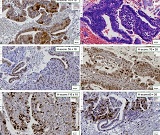
Benign/reactive lesions predominantly showed patchy or negative p16 staining patterns, wild-type p53 expression, and low proliferative activity, with Ki67 indices of 5% to 6%. In contrast, all precancerous lesions showed diffuse block-type p16 positivity and p53 overexpression. Ki67 proliferative activity was significantly increased in precancerous lesions, from 12% to 20% (p<0.001). The mean patient age was higher in precancerous lesions compared with benign/reactive lesions. HPV positivity was more frequently associated with precancerous lesions. Combined evaluation of p16, p53, and Ki67 revealed a clear immunophenotypic distinction between benign/reactive and precancerous glandular lesions.
Downloads
Gogitidze, G.-J., Kepuladze, S., & Burkadze, G. (2025). Features of Neural Microenvironment Remodelling in cervical intraepithelial neoplasia and squamous Cell Carcinoma. Georgian Scientists, 7(2), 96–109. https://doi.org/10.52340/gs.2025.07.02.09
Gogitidze, G., Kepuladze, S., Tevzadze, N., & Burkadze, G. (2023). The Role of the Local Neural microenvironment in the Progression of cervical intraepithelial Neoplasia. Georgian Scientists, 5(3), 182–188. https://doi.org/10.52340/2023.05.03.19
Gogitidze, G.-J., Kepuladze, S., & Burkadze, G. (2025). Vascular Remodeling Across the Spectrum of Cervical Intraepithelial Neoplasia and Squamous Cell Carcinoma: A Digital Pathology-Based Morphometric and Immunohistochemical Analysis. Georgian Scientists, 7(2), 56–68. https://doi.org/10.52340/gs.2025.07.02.06
Adamashvili, N., Beriashvili, R., Tevzadze, N., Kepuladze, S., & Burkadze, G. (2024). Features of the distribution of acute and chronic inflammatory index in cervical intraepithelial neoplasia of different degrees and its relationship with proliferative activity detected by AgNOR technology. Georgian Scientists, 6(1), 45–57. https://doi.org/10.52340/gs.2024.06.01.08
Pirog EC, Kleter B, Olgac S, et al. Prevalence of human papillomavirus DNA in different histological subtypes of cervical adenocarcinoma. American Journal of Pathology 2000;157(4):1055–1062.
Stolnicu S, Talia KL, McCluggage WG. The Evolving Spectrum of Precursor Lesions of Cervical Adenocarcinomas. Adv Anat Pathol 2020;27(5):278–293.
Makino H, Sato S, Yajima A, Komatsu S, Fukao A. Evaluation of the effectiveness of cervical cancer screening: a case-control study in Miyagi, Japan. Tohoku J Exp Med 1995;175(3):171–178.
Boria F, Siegrist J, Hardisson D, Saturio N, Zapardiel I. Lobular endocervical glandular hyperplasia mimicking cervical adenocarcinoma. J Obstet Gynaecol (Lahore) 2021;41(7):1166–1168.
Bosch FX, Burchell AN, Schiffman M, et al. Epidemiology and Natural History of Human Papillomavirus Infections and Type-Specific Implications in Cervical Neoplasia. Vaccine 2008;26(SUPPL. 10).
Nara M, Hashi A, Murata SI, et al. Lobular endocervical glandular hyperplasia as a presumed precursor of cervical adenocarcinoma independent of human papillomavirus infection. Gynecol Oncol 2007;106(2):289–298.
Liao, S. Y., Rodgers, W. H., Kauderer, J., Darcy, K. M., Carter, R., Susumu, N., Nagao, S., Walker, J. L., Hatae, M., & Stanbridge, E. J. (2013). Endocervical glandular neoplasia associated with lobular endocervical glandular hyperplasia is HPV-independent and correlates with carbonic anhydrase-IX expression: a Gynaecological Oncology Group Study. British journal of cancer, 108(3), 613–620. https://doi.org/10.1038/bjc.2012.578
Endocervical glandular neoplasia associated with lobular endocervical glandular hyperplasia is HPV-independent and correlates with carbonic anhydrase-IX expression: a Gynaecological Oncology Group Study - PMC [Homepage on the Internet]. [cited 2026 May 19];Available from: https://pmc.ncbi.nlm.nih.gov/articles/PMC3593541/
Yamazaki H, Sasagawa T, Basha W, Segawa T, Inoue M. Hybrid capture-II and LCR-E7 PCR assays for HPV typing in cervical cytologic samples. Int J Cancer 2001;94(2):222–227.
Liao SY, Rodgers WH, Kauderer J, et al. Carbonic anhydrase IX (CA-IX) and high-risk human papillomavirus (H-HPV) as diagnostic biomarkers of cervical dysplasia/neoplasia in Japanese women with a cytologic diagnosis of atypical glandular cells (AGC): A Gynecologic Oncology Group (GOG) Study. Br J Cancer 2011;104(2):353–360.
Klaes R, Friedrich T, Spitkovsky D, et al. Overexpression of p16ink4a as a specific marker for dysplastic and neoplastic epithelial cells of the cervix uteri. Int J Cancer 2001;92(2):276–284.
Negri G, Egarter-Vigl E, Kasal A, Romano F, Haitel A, Mian C. P16INK4a is a useful marker for the diagnosis of adenocarcinoma of the cervix uteri and its precursors: An immunohistochemical study with immunocytochemical correlations. American Journal of Surgical Pathology 2003;27(2):187–193.
Mehdi HK, Raju K, Sheela SR. Association of P16, Ki-67, and CD44 expression in high-grade squamous intraepithelial neoplasia and squamous cell carcinoma of the cervix. J Cancer Res Ther 2023;19(8):S260–S267.
Benevolo M, Mottolese M, Marandino F, et al. Immunohistochemical expression of p16INK4a is predictive of HR-HPV infection in cervical low-grade lesions. Modern Pathology 2006;19(3):384–391.
Nucci MR. Pseudoneoplastic glandular lesions of the uterine cervix: A selective review. International Journal of Gynecological Pathology 2014;33(4):330–338.
Gilks CB, Young RH, Aguirre P, DeLellis RA, Scully RE. Adenoma malignum (minimal deviation adenocarcinoma) of the uterine cervix. A clinicopathological and immunohistochemical analysis of 26 cases. American Journal of Surgical Pathology 1989;13(9):717–729.
M Fischer MQLSKE. The p53-p21-DREAM-CDE/CHR pathway regulates G2/M cell cycle genes. Nucleic Acids Res 2016;44:164–174.
Blagih J, Buck MD, Vousden KH. p53, cancer and the immune response. J Cell Sci 2020;133(5).
Copyright (c) 2026 Georgian Scientists

This work is licensed under a Creative Commons Attribution-NonCommercial-NoDerivatives 4.0 International License.