Synovial Membrane Cellular-molecular Profile, Vascular Changes in Rheumatoid Arthritis, Possible Markers of Disease Progression and Individual Treatment

Downloads


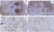
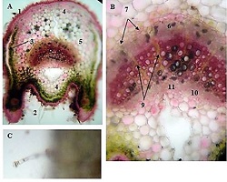
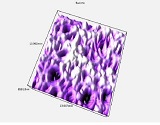
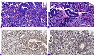
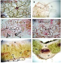
Rheumatoid arthritis is a chronic, progressive autoimmune disease characterized by persistent synovial inflammation and progressive joint destruction. The synovial membrane represents the primary target tissue of the disease, where complex cellular and molecular alterations contribute to chronic inflammation, tissue remodeling, and structural damage. Recent advances have demonstrated that rheumatoid synovium is a highly heterogeneous microenvironment composed of fibroblast-like synoviocytes, macrophages, T and B lymphocytes, endothelial cells, and multiple cytokine and chemokine signaling pathways. These interactions play a critical role in disease progression, angiogenesis, cartilage destruction, and variability in therapeutic response.
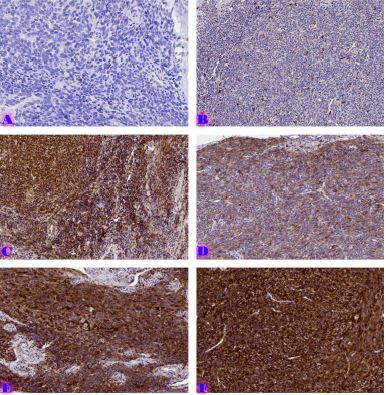
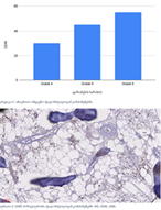
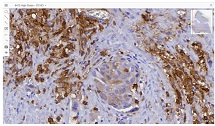
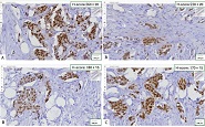
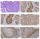
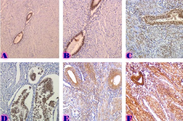
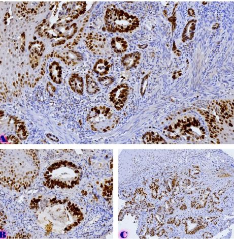
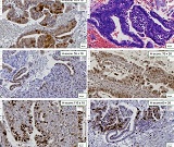
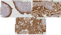
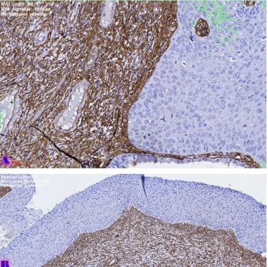
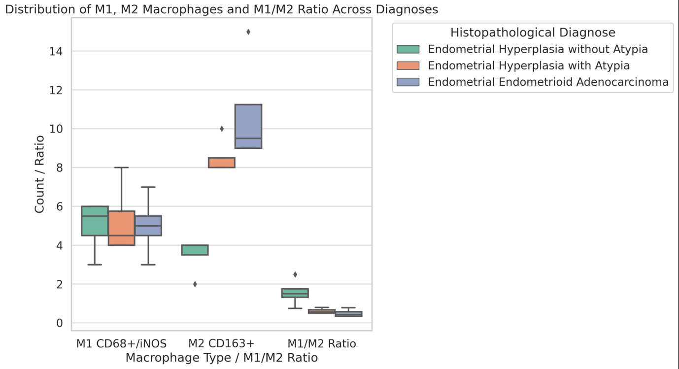
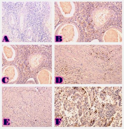
Increasing attention has been directed toward the histological and immunohistochemical characterization of synovial tissue. Biomarkers such as CD64, CD68, CD163, VEGF, CXCL13, and ICAM-1 reflect inflammatory activity, macrophage polarization, angiogenic potential, and stromal remodeling within the synovial microenvironment. Moreover, distinct synovial histopathological phenotypes, including lymphoid, myeloid, pauci-immune, and fibroid patterns, may provide important prognostic and predictive information for individualized therapeutic strategies.
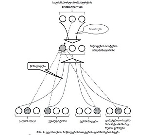
The development of synovial biopsy techniques, digital pathology, and advanced molecular technologies has significantly expanded the potential of personalized rheumatology. Detailed evaluation of the cellular-molecular profile and vascular alterations of the synovial membrane may improve understanding of rheumatoid arthritis pathogenesis and contribute to the identification of prognostic biomarkers and targeted therapeutic approaches.
Downloads
Yamanishi Y, Firestein GS. Pathogenesis of rheumatoid arthritis: The role of synoviocytes. Rheumatic Disease Clinics of North America 2001;27:355–71. https://doi.org/10.1016/S0889-857X(05)70206-4.
Cooles FAH, Isaacs JD, Anderson AE. Treg cells in rheumatoid arthritis: An update. Curr Rheumatol Rep 2013;15. https://doi.org/10.1007/S11926-013-0352-0.
Müller-Ladner U, Ospelt C, Gay S, Distler O, Pap T. Cells of the synovium in rheumatoid arthritis. Synovial fibroblasts. Arthritis Res Ther 2007;9. https://doi.org/10.1186/AR2337.
Schellekens GA, De Jong BAW, Van Den Hoogen FHJ, Van De Putte LBA, Van Venrooij WJ. Citrulline is an essential constituent of antigenic determinants recognized by rheumatoid arthritis-specific autoantibodies. Journal of Clinical Investigation 1998;101:273–81. https://doi.org/10.1172/JCI1316.
Put S, Westhovens R, Lahoutte T, Matthys P. Molecular imaging of rheumatoid arthritis: Emerging markers, tools, and techniques. Arthritis Res Ther 2014;16. https://doi.org/10.1186/AR4542.
Haynes DR, Barg E, Crotti TN, Holding C, Weedon H, Atkins GJ, et al. Osteoprotegerin expression in synovial tissue from patients with rheumatoid arthritis, spondyloarthropathies and osteoarthritis and normal controls. Rheumatology 2003;42:123–34. https://doi.org/10.1093/RHEUMATOLOGY/KEG047.
Klaasen R, Thurlings RM, Wijbrandts CA, Van Kuijk AW, Baeten D, Gerlag DM, et al. The relationship between synovial lymphocyte aggregates and the clinical response to infliximab in rheumatoid arthritis: A prospective study. Arthritis Rheum 2009;60:3217–24. https://doi.org/10.1002/ART.24913.
Klaasen R, Thurlings RM, Wijbrandts CA, Van Kuijk AW, Baeten D, Gerlag DM, et al. The relationship between synovial lymphocyte aggregates and the clinical response to infliximab in rheumatoid arthritis: A prospective study. Arthritis Rheum 2009;60:3217–24. https://doi.org/10.1002/ART.24913.
Lequerré T, Bansard C, Vittecoq O, Derambure C, Hiron M, Daveau M, et al. Early and long-standing rheumatoid arthritis: Distinct molecular signatures identified by gene-expression profiling in synovia. Arthritis Res Ther 2009;11. https://doi.org/10.1186/AR2744.
Mor A, Abramson SB, Pillinger MH. The fibroblast-like synovial cell in rheumatoid arthritis: A key player in inflammation and joint destruction. Clinical Immunology 2005;115:118–28. https://doi.org/10.1016/J.CLIM.2004.12.009.
Kinne RW, Bräuer R, Stuhlmüller B, Palombo-Kinne E, Burmester GR. Macrophages in rheumatoid arthritis. Arthritis Res 2000;2:189–202. https://doi.org/10.1186/AR86.
Ling S, Cline EN, Haug TS, Fox DA, Holoshitz J. Citrullinated calreticulin potentiates rheumatoid arthritis shared epitope signaling. Arthritis Rheum 2013;65:618–26. https://doi.org/10.1002/ART.37814.
Yoshida K, Korchynskyi O, Tak PP, Isozaki T, Ruth JH, Campbell PL, et al. Citrullination of Epithelial Neutrophil-Activating Peptide 78/CXCL5 Results in Conversion from a Non-Monocyte-Recruiting Chemokine to a Monocyte-Recruiting Chemokine. Arthritis and Rheumatology 2014;66:2716–27. https://doi.org/10.1002/ART.38750.
McInnes IB, Schett G. Cytokines in the pathogenesis of rheumatoid arthritis. Nat Rev Immunol 2007;7:429–42. https://doi.org/10.1038/NRI2094.
Asif Amin M, Fox DA, Ruth JH. Synovial Cellular and Molecular Markers in Rheumatoid Arthritis. Semin Immunopathol 2017;39:385. https://doi.org/10.1007/S00281-017-0631-3.
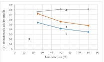
Qavtaradze N, Qartvelishvili E (Dodo), Kepuladze S, Burkadze G. Structural-molecular features of the osteochondral unit of the femur head and the synovial membrane and characteristics of progression and relapse in different types of osteoarthritis. Georgian Scientists 2022;4:175–84. https://doi.org/10.52340/gs.2022.04.05.18.
Germisashvili K, Kartvelisvhili E, Burkadze G, Kepuladze S, Kantaria N. Proliferative-apoptotic, hormonal receptor, and immune response characteristics of rheumatoid arthritis. Georgian Scientists 2023;5:66–74. https://doi.org/10.52340/2023.05.03.08.
Stephenson W, Donlin LT, Butler A, Rozo C, Bracken B, Rashidfarrokhi A, et al. Single-cell RNA-seq of rheumatoid arthritis synovial tissue using low-cost microfluidic instrumentation. Nat Commun 2018;9. https://doi.org/10.1038/S41467-017-02659-X.
Orange DE, Agius P, DiCarlo EF, Robine N, Geiger H, Szymonifka J, et al. Identification of Three Rheumatoid Arthritis Disease Subtypes by Machine Learning Integration of Synovial Histologic Features and RNA Sequencing Data. Arthritis and Rheumatology 2018;70:690–701. https://doi.org/10.1002/ART.40428.
Orange DE, Agius P, DiCarlo EF, Robine N, Geiger H, Szymonifka J, et al. Identification of Three Rheumatoid Arthritis Disease Subtypes by Machine Learning Integration of Synovial Histologic Features and RNA Sequencing Data. Arthritis and Rheumatology 2018;70:690–701. https://doi.org/10.1002/ART.40428.
Bartok B, Firestein GS. Fibroblast-like synoviocytes: Key effector cells in rheumatoid arthritis. Immunol Rev 2010;233:233–55. https://doi.org/10.1111/J.0105-2896.2009.00859.X.
Gordon S, Martinez FO. Alternative activation of macrophages: Mechanism and functions. Immunity 2010;32:593–604. https://doi.org/10.1016/J.IMMUNI.2010.05.007.
Koch AE, Polverini PJ, Kunkel SL, Harlow LA, DiPietro LA, Elner VM, et al. Interleukin-8 as a Macrophage-Derived Mediator of Angiogenesis. Science (1979) 1992;258:1798–801. https://doi.org/10.1126/SCIENCE.1281554.
Strieter RM, Polverini PJ, Kunkel SL, Arenberg DA, Burdick MD, Kasper J, et al. The functional role of the ELR motif in CXC chemokine-mediated angiogenesis. Journal of Biological Chemistry 1995;270:27348–57. https://doi.org/10.1074/JBC.270.45.27348.
Yamada R, Sano H, Hla T, Hashiramoto A, Kawahito Y, Mukai S, et al. Selective inhibition of cyclooxygenase-2 with antisense oligodeoxynucleotide restricts induction of rat adjuvant-induced arthritis. Biochem Biophys Res Commun 2000;269:415–21. https://doi.org/10.1006/BBRC.2000.2303.
Cauli A, Yanni G, Panayi GS. Interleukin-1, interleukin-1 receptor antagonist and macrophage populations in rheumatoid arthritis synovial membrane. Br J Rheumatol 1997;36:935–40. https://doi.org/10.1093/RHEUMATOLOGY/36.9.935.
Xing R, Jin Y, Sun L, Yang L, Li C, Li Z, et al. Interleukin-21 induces migration and invasion of fibroblast-like synoviocytes from patients with rheumatoid arthritis. Clin Exp Immunol 2016;184:147–58. https://doi.org/10.1111/CEI.12751.
Hashimoto A, Tarner IH, Bohle RM, Gaumann A, Manetti M, Distler O, et al. Analysis of vascular gene expression in arthritic synovium by laser-mediated microdissection. Arthritis Rheum 2007;56:1094–105. https://doi.org/10.1002/ART.22450.
Audoy-Rémus J, Richard JF, Soulet D, Zhou H, Kubes P, Vallières L. Rod-shaped monocytes patrol the brain vasculature and give rise to perivascular macrophages under the influence of proinflammatory cytokines and angiopoietin-2. Journal of Neuroscience 2008;28:10187–99. https://doi.org/10.1523/JNEUROSCI.3510-08.2008.
Hashimoto A, Tarner IH, Bohle RM, Gaumann A, Manetti M, Distler O, et al. Analysis of vascular gene expression in arthritic synovium by laser-mediated microdissection. Arthritis Rheum 2007;56:1094–105. https://doi.org/10.1002/ART.22450.
Gerlag DM, Tak PP. How to perform and analyse synovial biopsies. Best Pract Res Clin Rheumatol 2013;27:195–207. https://doi.org/10.1016/J.BERH.2013.03.006.
Pitzalis C, Kelly S, Humby F. New learnings on the pathophysiology of RA from synovial biopsies. Curr Opin Rheumatol 2013;25:334–44. https://doi.org/10.1097/BOR.0B013E32835FD8EB.
Pitzalis C, Kelly S, Humby F. New learnings on the pathophysiology of RA from synovial biopsies. Curr Opin Rheumatol 2013;25:334–44. https://doi.org/10.1097/BOR.0B013E32835FD8EB.
Bresnihan B, Tak PP, Emery P, Klareskog L, Breedveld F. Synovial biopsy in arthritis research: Five years of concerted European collaboration. Ann Rheum Dis 2000;59:506–10. https://doi.org/10.1136/ARD.59.7.506.
Copyright (c) 2026 Georgian Scientists

This work is licensed under a Creative Commons Attribution-NonCommercial-NoDerivatives 4.0 International License.