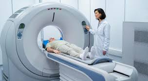
Diagnostic Criteria for Complicated Diverticular Disease of the Colon Using Contrast-Enhanced Computed Tomography

Despite extensive research and publications on diverticular disease of the colon (DDC), the diagnostic algorithm and treatment strategy remain subjects of ongoing debate. The incidence of DDC is steadily increasing. The age distribution of patients diagnosed with complicated and uncomplicated DDC is as follows: under 30 years – less than 1%, under 40 years – 5%, under 60 years – 30%, and over 80 years – 50-60%. Approximately 10-25% of patients develop diverticulitis, and 15% of them experience a more severe complication – perforation with peritonitis. Our study aimed to develop diagnostic criteria for complications of diverticular disease of the colon (DDC) using contrast-enhanced computed tomography (CT) for determining treatment tactics. Methodically we analyzed 128 CT scans with intravenous contrast performed on patients with various manifestations of DDC complications, who were treated at the proctology department of the Minsk Regional Clinical Hospital between January 2020 and December 2023. Bowel preparation was carried out using a special diet and laxatives containing polyethylene glycol. A risk assessment scale was developed and four risk groups were formed, to which patients were assigned depending on the number of points: 0 points - no risk, 1-3 points - low risk, 4-5 points - moderate risk, 6 or more points – high risk. According to the results of our study, we determined the extent of diverticula distribution along the segments of the colon: isolated sigmoid - 71 (55.5%), left colon - 38 (29.7%), right colon - 1 (0.8%), total involvement - 18 (14%). Establish the localization of diverticula in relation to the mesentery was established: mesenteric border – 67 (52.3%), anti-mesenteric (free) border - 23 (18%), mixed localization (mesenteric and anti-mesenteric) - 38 (29.7%). The analysis of the obtained results allowed us to determine the treatment tactics for each patient depending on the nature of the existing complications of diverticular disease of the colon. Using a diagnostic map during computed tomography with bolus contrast is key for developing diagnostic criteria for complications of diverticular colon disease. Diagnostic maps are essential for accurate diagnosis and preoperative treatment planning.
Downloads
Metrics
No metrics found.
Vermeulen J., Lange J.F. Treatment of perforated diverticulitis with generalized peritonitis: past, present, and future. World J Surg. 2010;34(3):587-593. doi: 10.1007/s00268-009-0372-0.
Shaikh S., Krukowski Z.H. Outcome of a conservative policy for managing acute sigmoid diverticulitis. Br J Surg. 2007;94(7):876-879. doi: 10.1002/bjs.5703.
Ivashkin V.T., Shelygin Yu.A., Achkasov S.I., Vasilyev S.V., Grigoryev Ye.G., Dudka V.V., et al. Diagnostics and treatment of diverticular disease of the colon: guidelines of the Russian gastroenterological Association and Russian association of coloproctology. Rus J Gastroenterol Hepatol Coloproctol. 2016;26(1):65-80. doi.org/10.22416/1382-4376-2016-26-1-65-80.
Gielens M.P., Mulder I.M., van der Harst E., Gosselink M.P., Kraal K.J., Teng H.T., et al. Preoperative staging of perforated diverticulitis by computed tomography scanning. Tech Coloproctol. 2012;16(5):363-368. doi: 10.1007/s10151-012-0853-2.
Kaiser A.M., Jiang J.K., Lake J.P., Ault G., Artinyan A., Gonzalez-Ruiz C. et al. The management of complicated diverticulitis and the role of computed tomography. Am J Gastroenterol. 2005;100(4):910-917. doi: 10.1111/j.1572-0241.2005.41154.x.
Krukowski Z.H., Matheson N.A. Emergency surgery for diverticular disease complicated by generalized and faecal peritonitis: a review. Br J Surg. 1984;71(12):921-927. doi: 10.1002/bjs.1800711202.
Lammers B.J., Schumpelick V., Röher H.D. Standards in der Diagnostik der Divertikulitis. Chirurg. 2002;73(7):670-674. doi: 10.1007/s00104-002-0496-3.
Laméris W., van Randen A., van Gulik T.M., Busch O.R., Winkelhagen J., Bossuyt P.M., et al. A clinical decision rule to establish the diagnosis of acute diverticulitis at the emergency department. Dis Colon Rectum. 2010;53(6):896-904. doi: 10.1007/DCR.0b013e3181d98d86.
Tack D., Bohy P., Perlot I., De Maertelaer V., Alkeilani O., Sourtzis S., et al. Suspected acute colon diverticulitis: imaging with low-dose unenhanced multi-detector row CT. Radiology. 2005;237(1):189-196. doi: 10.1148/radiol.2371041432.
McGillicuddy E.A., Schuster K.M., Davis K.A., Longo W.E. Factors predicting morbidity and mortality in emergency colorectal procedures in elderly patients. Arch Surg. 2009;144(12):1157-1162. doi: 10.1001/archsurg.2009.203.
Shelygin Yu.A., Achkasov S.I., Moskalev A.I. Classification of diverticular disease. Koloproktologia. 2014;4:5-13.
Hadji-Ismail I.A., Rummo O.O., Varabei A.V., Senkevich O.I., Marakhouskaja E.I. Rare localization of diverticula of the colon. Proceedings of the National Academy of Sciences of Belarus, Medical series. 2023;20(1):28-33. (In Russ.) https://doi.org/10.29235/1814-6023-2023-20-1-28-33.
Hadji -Ismail I.A. Classification of diverticular colon disease. Healthcare. 2022;5:21-28.
Neff C.C., van Sonnenberg E. CT of diverticulitis. Diagnosis and treatment. Radiol Clin North Am. 1989;27(4):743-752.
Hawkins A.T., Wise P.E., Chan T., Lee J.T., Glyn T., Wood V., et al. Diverticulitis: an update from the age old paradigm. Curr Probl Surg. 2020;57(10):100862. doi: 10.1016/j.cpsurg.2020.100862.
Sartelli M., Moore F.A., Ansaloni L., Di Saverio S., Coccolini F., Griffiths E.A., et al. A proposal for a CT driven classification of left colon acute diverticulitis. World J Emerg Surg. 2015;10:3. doi: 10.1186/1749-7922-10-3.
Poletti P.A., Platon A., Rutschmann O., Kinkel K., Nyikus V., Ghiorghiu S., et al. Acute left colonic diverticulitis: can CT findings be used to predict recurrence? AJR Am J Roentgenol. 2004;182(5):1159-1165. doi: 10.2214/ajr.182.5.1821159.
Copyright (c) 2025 Georgian Scientists

This work is licensed under a Creative Commons Attribution-NonCommercial-NoDerivatives 4.0 International License.


