ასაკისა და რეფრაქციის კორელაცია ბადურისა და ქორიოიდეის სისქესთან ახლომხედველ ბავშვებში
ჩამოტვირთვები
ახლომხედველ თვალებში ქორიოიდეასა და ბადურისმორფოლოგიური ცვლილებები კარგადაა შესწავლილი მოზრდილებში, მაგრამ ეს ცვლილებები არასაკმარისადაა აღწერილი ბავშვებში. კვლევები, სადაც აღწერილია ბადურის და ქორიოიდეის სისქე სხვადასხვა რეფრაქციული მდგომარეობის მქონე ბავშვებში მცირერიცხოვანია.
კვლევის მიზანს წარმოადგენდა ასაკისა და რეფრაქციის კორელაციის შეფასება ბადურისა და ქორიოიდეის სისქესთან ახლომხედველ ბავშვებში SS-OCT-ის გამოყენებით.
ჩატარებულია ჯვარედინი კვლევა ქორიოიდეისა დაბადურის შრეების ანატომიური და ტოპოგრაფიული ვარიაციების გასარკვევად ახლომხედველ ბავშვებში. გამოყენებულია SSOCT (SS-OCTA DRI Triton Topcon) მოწყობილობა ფართოზოლიაინ ინფრაწითელთან მიახლოებული სუპერლუმინესცენციური დიოდით, რომლის სინათლის წყაროს ტალღის სიგრძე 1050 ნმ-ია, ასევე, ერთჯერადი ფოტოდიოდი, როგორც დეტექტორი. SS-OCTA აწყობილია შემოწმებულ კლინიკურ პლატფორმაზე DRI Triton ეფუძნება CTARA-ს დაპატენტებულ ალგორითმს.
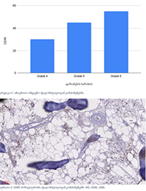
კვლევის მონაცემების მიხედვით, ქორიოიდეისა და ბადურის სისქე მჭიდროდ კორელირებს SER-თან და AL-თან. ბადურის პერიფოვეალური სისქე მიოპიის მქონე ბავშვებში უფრო მცირეა, ვიდრე ემეტროპებში. წინამორბედი კვლევების კონტექსტში, სადაც ავტორები ვარაუდობენ, რომ ქორიოიდეის მიდამოში გამოვლენილი ცვლილებებიწინ უსწრებს სკლერის ცვლილებებს ინდუცირებულ ამეტროპიაში, ავტორები თვლიან, რომ მიოპიის პროგრესირების ადრეულ სტადიაზე ვითარდება ჯერ ქორიოიდის გათხელება, რასაც მოსდევს ბადურის გათხელება პერიფოვეალურ მიდამოში, რაც პროგრესირებს ცენტრიდანული მიმართულებით. კვლევის შედეგების ინტერპრეტაციისას აუცილებლად მხედველობაშია მისაღები ის, რომ SSOCT-ით გაზომილი ქორიოიდეის სისქე მეტია, ვიდრე სხვა OCT ინსტრუმენტების გამოყენების შემთხვევაში. ამრიგად, კვლევის ქორიოიდეის სისქის მონაცემები არ შეიძლება პირდაპირ შედარდეს სხვა OCT ინსტრუმენტების გამოყენებით ჩატარებული კვლევების მონაცემებს.
დადგენილია, რომ ახლომხედველ ბავშვებს აქვთ უფრო თხელი ქორიოიდეა ყველა მიდამოში და უფრო თხელი ბადურა ზედა და ქვედა პერიფოვეალურ რეგიონებში, ემეტროპებთან შედარებით. ქორიოიდეის სისქე ფოვეის მიდამოში მჭიდროდ კორელირებს აქსიალურ სიგრძესადა რეფრაქციის ხარისხთან.
Downloads
Dolgin E. The myopia boom. Nature 2015;519(7543):276–278.
Morgan IG, Ohno-Matsui K, Saw SM. Myopia. Lancet 2012;379(9827):1739–1748.
Cook RC, Glasscock RE. Refractive and ocular findings in thenewborn. Am J Ophthalmol 1951;34(10):1407–1413.
Summers JA. The choroid as a sclera growth regulator. Exp Eye Res 2013;114:120–127.
Vincent SJ, Collins MJ, Read SA, Carney LG. Retinal and choroidal thickness in myopic an isometropia. Invest Ophthalmol Vis Sci 2013;54(4):2445–2456.
Nickla DL, Wildsoet C, Wallman J. Compensation for spectacle lenses involves changes in proteoglycan synthesis in both the sclera and choroid. Curr Eye Res 1997; 16(4):320–326
Wallman J, Wildsoet C, Xu A, et al. Moving the retina: choroidal modulation of refractive state. Vision Res 1995;35(1):37–50.
Wildsoet C, Wallman J. Choroidal and scleral mechanisms of compensation for spectacle lenses in chicks. Vision Res 1995;35(9):1175–1194.
Wildsoet CF. Active emmetropization-evidence for its existence and ramifications for clinical practice. Ophthalmic Physiol Opt. 1997;17(4):279-290.
Wong AC, Chan CW, Hui SP. Relationship of gender, body mass index, and axial length with central retinal thickness using optical coherence tomography. Eye 2005;19(3):292–297.
Lim MC, Hoh ST, Foster PJ, et al. Use of optical coherence tomography to assess variations in macular retinal thickness in myopia. Invest Ophthalmol Vis Sci 2005;46(3):974–978.
Yamashita T, Tanaka M, Kii Y, Nakao K, Sakamoto T. Association between retinal thickness of 64 sectors in posterior pole determined by optical coherence tomography and axial length and body height. Invest Ophthalmol. Vis Sci 2013;54(12):7478–7482.
Pang Y, Goodfellow GW, Allison C, Block S, Frantz KA. A prospective study of macular thickness in amblyopic children with unilateral high myopia. Invest Ophthalmol Vis Sci 2011; 52(5):2444–2449.
Lam DS, Leung KS, Mohamed S, et al. Regional variations in the relationship between macular thickness measurements and myopia. Invest Ophthalmol Vis Sci 2007;48(1):376–382.
Luo HD, Gazzard G, Fong A, et al. Myopia, axial length, and OCT characteristics of the macula in Singaporean children. Invest Ophthalmol Vis Sci 2006;47(7): 2773–2781.
Nishida Y, Fujiwara T, Imamura Y, Lima LH, Kurosaka D,Spaide RF. Choroidal thickness and visual acuity in highly myopic eyes. Retina 2012;32(7):1229–1236.
Read SA, Alonso-Caneiro D, Vincent SJ, Collins MJ. Longitudinal changes in choroidal thickness and eye growth in childhood. Invest Ophthalmol Vis Sci 2015;56(5):3103–3112.
Zhang Z, He X, Zhu J, Jiang K, Zheng W, K. B. Macular measurements using optical coherence tomography in healthy Chinese school age children. Invest Ophthalmol VisSci 2011;52(9):6377–6383.
Unterhuber A, Povazay B, Hermann B, Sattmann H, Chavez-Pirson A, Drexler W. In vivo retinal optical coherence tomography at 1040 nm - enhanced penetration into the choroid. Opt Express 2005;13(9):3252–3258.
Matsuo Y, Sakamoto T, Yamashita T, Tomita M, ShirasawaM, Terasaki H. Comparisons of choroidal thickness of normal eyes obtained by two different spectral- domain OCT instruments and one swept-source OCT instrument. Invest Ophthalmol Vis Sci 2013;54(12): 7630–7636.
Read SA, Collins MJ, Vincent SJ, Alonso-Caneiro D. Choroidal thickness in myopic and no myopic children assessed with enhanced depth imaging optical coherence tomography. Invest Ophthalmol Vis Sci 2013;54(12): 7578–7586.
Harb E, Hyman L, Gwiazda J, et al. Choroidal thickness profiles in myopic eyes of young adults in the Correction of Myopia Evaluation Trial Cohort. Am J Ophthalmol 2015; 160(1):62–71.e62.
Hirata M, Tsujikawa A, Matsumoto A, et al. Macular choroidal thickness and volume in normal subjects measured by swept-source optical coherence tomography. Invest Ophthalmol Vis Sci 2011;52(8):4971–4978.
Bidaut-Garnier M, Schwartz C, Puyraveau M, Montard M, Delbosc B, Saleh M. Choroidal thickness measurement in children using optical coherence tomography. Retina 2014; 34(4):768–774.
Ikuno Y, Tano Y. Retinal and choroidal biometry in highly myopic eyes with spectral domain optical coherence tomography. Invest Ophthalmol Vis Sci 2009;50(8):3876–3880.
Margolis R, Spaide RF. A pilot study of enhanced depth imaging optical coherence tomography of the choroid in normal eyes. Am J Ophthalmol 2009;147(5):811–815.
Ho M, Liu DT, Chan VC, Lam DS. Choroidal thickness measurement in myopic eyes by enhanced depth optical coherence tomography. Ophthalmology 2013;120(9):1909–1914.
Ikuno Y, Kawaguchi K, Nouchi T, Yasuno Y. Choroidal thickness in healthy Japanese subjects. Invest Ophthalmol Vis Sci 2010;51(4):2173–2176.
Fujiwara T, Imamura Y, Margolis R, Slakter JS, Spaide RF. Enhanced depth imaging optical coherence tomography of the choroid in highly myopic eyes. Am J Ophthalmol 2009; 148(3):445–450.
Shao L, Xu L, Wei WB, et al. Visual acuity and subfoveal choroidal thickness: the Beijing Eye Study. Am J Ophthalmol. 2014;158(4):702-709.e1. doi:10.1016/j.ajo.2014.05.023
Flores-Moreno I, Ruiz-Medrano J, Duker JS, Ruiz-Moreno JM. The relationship between retinal and choroidal thickness and visual acuity in highly myopic eyes. Br J Ophthalmol 2013; 97(8):1010–1013.
Al-Haddad C, El Chaar L, Antonios R, El-Dairi M, Noureddin B. Interocular symmetry in macular choroidal thickness in children. J Ophthalmol. 2014; 2014:472391. doi:10.1155/2014/472391
Karahan E, Zengin MO, Tuncer I. Correlation of choroidal thickness with outer and inner retinal layers. Ophthalmic Surg Lasers Imaging Retina 2013;44(6):544–548.
Chen S, Wang B, Dong N, Ren X, Zhang T, Xiao L. Macular measurements using spectral-domain optical coherence tomography in Chinese myopic children. Invest Ophthalmol. Vis Sci 2014;55(11):7410–7416.
Huang J, Liu X, Wu Z, Xiao H, Dustin L, Sadda S. Macular thickness measurements in normal eyes with time-domain and Fourier-domain optical coherence tomography. Retina2009;29(7):980–987.
Ooto S, Hangai M, Tomidokoro A, et al. Effects of age, sex, and axial length on the three-dimensional profile of normal macular layer structures. Invest Ophthalmol Vis Sci 2011; 52(12):8769–8779.
Song WK, Lee SC, Lee ES, Kim CY, Kim SS. Macular thickness variations with sex, age, and axial length in healthy subjects: a spectral domain-optical coherence tomography study. Invest Ophthalmol Vis Sci 2010;51(8):3913–3918.
Grossniklaus HE, Green WR. Pathologic findings in pathologic myopia. Retina 1992;12(2):127–133.
Wu PC, Chen YJ, Chen CH, et al. Assessment of macular retinal thickness and volume in normal eyes and highly myopic eyes with third-generation optical coherence tomography. Eye 2008;22(4):551–555.
Liu, X., Shen, M., Yuan, Y., Huang, S., Zhu, D., Ma, Q., Ye, X., & Lu, F. (2015). Macular Thickness Profiles of Intraretinal Layers in Myopia Evaluated by Ultrahigh-Resolution Optical Coherence Tomography. American journal of ophthalmology, 160(1), 53–61.e2. https://doi.org/10.1016/j.ajo.2015.03.012
Ooto S, Hangai M, Sakamoto A, et al. Three-dimensional profile of macular retinal thickness in normal Japanese eyes. Invest Ophthalmol Vis Sci 2010;51(1):465–473.
Kim, N. R., Kim, J. H., Lee, J., Lee, E. S., Seong, G. J., & Kim, C. Y. (2011). Determinants of perimacular inner retinal layer thickness in normal eyes measured by Fourier-domain optical coherence tomography. Investigative ophthalmology & visual science, 52(6), 3413–3418. https://doi.org/10.1167/iovs.10-6648
Kim, M. J., Lee, E. J., & Kim, T. W. (2010). Peripapillary retinal nerve fibre layer thickness profile in subjects with myopia measured using the Stratus optical coherence tomography. The British journal of ophthalmology, 94(1), 115–120. https://doi.org/10.1136/bjo.2009.162206
Kang, S. H., Hong, S. W., Im, S. K., Lee, S. H., & Ahn, M. D. (2010). Effect of myopia on the thickness of the retinal nerve fiber layer measured by Cirrus HD optical coherence tomography. Investigative ophthalmology & visual science, 51(8), 4075–4083. https://doi.org/10.1167/iovs.09-4737
Zhu BD, Li SM, Li H, et al. Retinal nerve fiber layer thickness in a population of 12-year-old children in central China measured by iVue-100 spectral-domain optical coherence tomography: the Anyang Childhood Eye Study. Invest Ophthalmol Vis Sci 2013;54(13):8104–8111.
Leung, C. K., Mohamed, S., Leung, K. S., Cheung, C. Y., Chan, S. L., Cheng, D. K., Lee, A. K., Leung, G. Y., Rao, S. K., & Lam, D. S. (2006). Retinal nerve fiber layer measurements in myopia: An optical coherence tomography study. Investigative ophthalmology & visual science, 47(12), 5171–5176. https://doi.org/10.1167/iovs.06-0545
Hoh, S. T., Lim, M. C., Seah, S. K., Lim, A. T., Chew, S. J., Foster, P. J., & Aung, T. (2006). Peripapillary retinal nerve fiber layer thickness variations with myopia. Ophthalmology, 113(5), 773–777. https://doi.org/10.1016/j.ophtha.2006.01.058
საავტორო უფლებები (c) 2023 ქართველი მეცნიერები

ეს ნამუშევარი ლიცენზირებულია Creative Commons Attribution-NonCommercial-NoDerivatives 4.0 საერთაშორისო ლიცენზიით .


