The Role of M1 and M2 Macrophages in the Progression of Endometrial Hyperplasia to Endometrioid Adenocarcinoma
Загрузки
Background: The progression of endometrial pathologies, from benign hyperplasia to malignant adenocarcinoma, involves complex interactions between the immune microenvironment, hormonal receptor dynamics, and systemic conditions. This study aims to evaluate immune markers, such as M1/M2 macrophage ratios and tumour-infiltrating lymphocytes (TILs), alongside estrogen (ER) and progesterone (PR) receptor expression in different endometrial conditions.
Methods: This retrospective study included 120 cases categorised into three groups: endometrial hyperplasia without atypia, hyperplasia with atypia, and endometrial endometrioid adenocarcinoma. Immune markers (M1/M2 ratio, TILs) and hormonal receptors (ER, PR) were assessed using immunohistochemistry. Statistical comparisons were conducted across diagnostic groups stratified by age and systemic conditions.
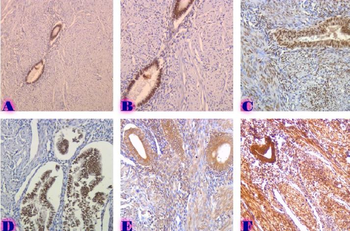
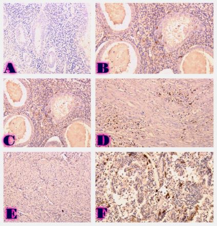
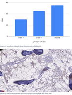
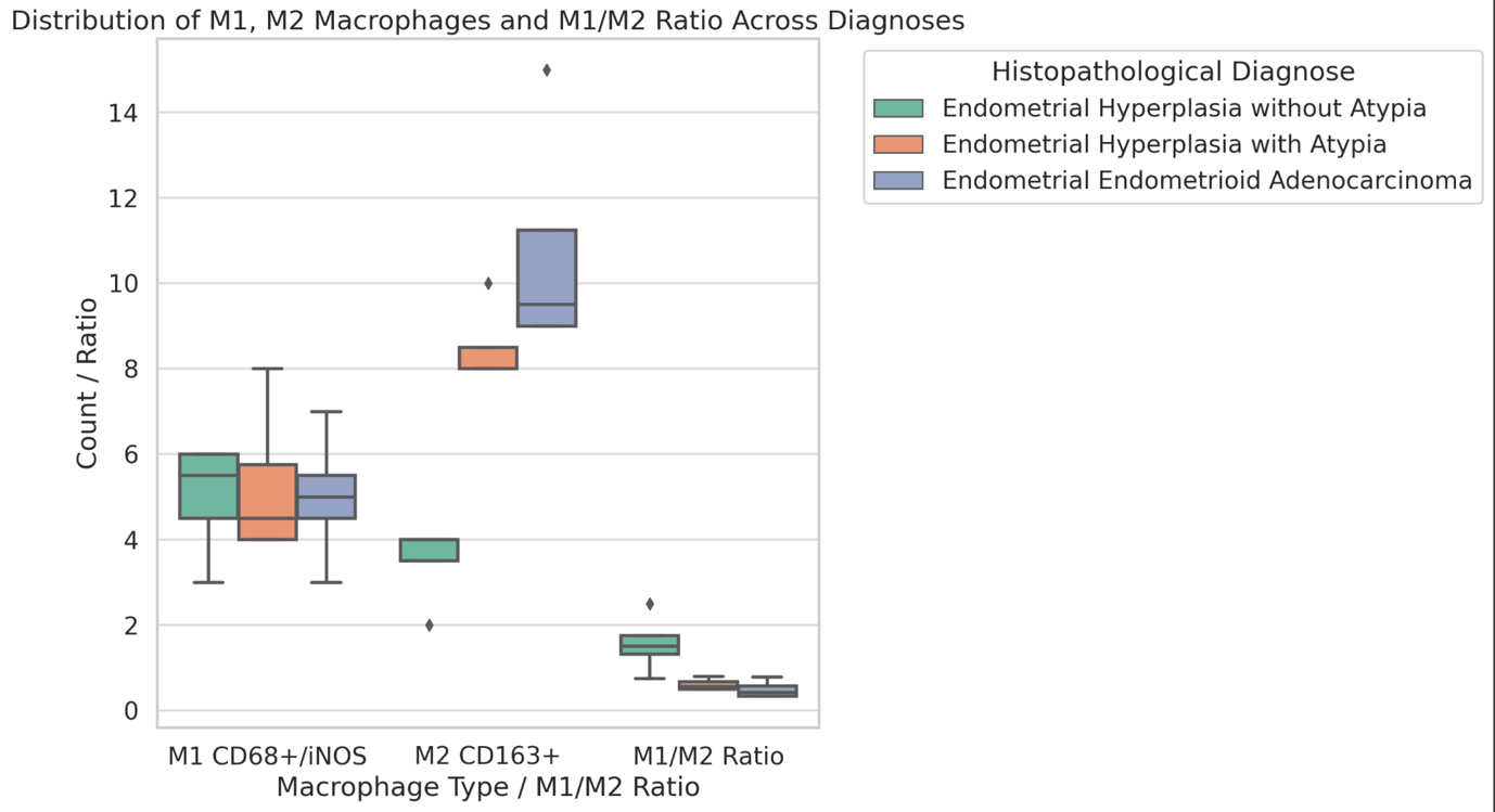
Results: The M1/M2 Ratio declined progressively from hyperplasia without atypia (mean: 2.0) to adenocarcinoma (mean: 0.5, p < 0.01), indicating a shift from a pro-inflammatory to an immunosuppressive microenvironment. TILs %: Minimal in hyperplasia without atypia (0%), increasing significantly in adenocarcinoma (mean: 15%, p < 0.01). ER/PR expression decreased from hyperplasia without atypia (ER: 80%, PR: 70%) to adenocarcinoma (ER: 30%, PR: 20%, p < 0.01). Premenopausal women exhibited higher receptor expression than postmenopausal women (p < 0.05).
Conclusion: This study demonstrates the critical role of immune and hormonal markers in the progression of endometrial diseases. The decline in M1/M2 ratios, rising TILs, and reduced ER/PR expression correlate with disease severity and systemic conditions. These findings suggest the potential of targeting the immune microenvironment and hormonal pathways for personalised therapeutic strategies. Future research should explore the molecular mechanisms underlying these changes and validate these biomarkers in prospective studies.
Скачивания
J. Tu et al., “Growth arrest-specific transcript 5 represses endometrial cancer development by promoting antitumor function of tumor-associated macrophages,” Cancer Sci, vol. 113, no. 8, pp. 2496–2512, Aug. 2022, doi: 10.1111/cas.15390.
S. Mei et al., “PRMT5 promotes progression of endometrioid adenocarcinoma via ERα and cell cycle signaling pathways,” Journal of Pathology: Clinical Research, vol. 7, no. 2, pp. 154–164, Mar. 2021, doi: 10.1002/cjp2.194.
M. Bied, W. W. Ho, F. Ginhoux, and C. Blériot, “Roles of macrophages in tumor development: a spatiotemporal perspective,” Cell Mol Immunol, vol. 20, no. 9, pp. 983–992, Sep. 2023, doi: 10.1038/s41423-023-01061-6.
H. Tong et al., “Tumor-associated macrophage-derived CXCL8 could induce ERα suppression via HOXB13 in endometrial cancer,” Cancer Lett, vol. 376, no. 1, pp. 127–136, Jun. 2016, doi: 10.1016/J.CANLET.2016.03.036.
B. Allard, M. S. Longhi, S. C. Robson, and J. Stagg, “The ectonucleotidases CD39 and CD73: Novel checkpoint inhibitor targets,” Immunol Rev, vol. 276, no. 1, pp. 121–144, Mar. 2017, doi: 10.1111/IMR.12528.
A. S. Uduwela, M. A. K. Perera, L. Aiqing, and I. S. Fraser, “Endometrial-myometrial interface: Relationship to adenomyosis and changes in pregnancy,” Obstet Gynecol Surv, vol. 55, no. 6, pp. 390–400, Jun. 2000, doi: 10.1097/00006254-200006000-00025.
C. Ozturk, G. Askan, S. Duman Ozturk, O. Okcu, B. Sen, and R. Bedir, “High Tumor Infiltrating Lymphocytes Are Associated with Overall Survival and Good Prognostic Parameters in Endometrial Endometrioid Carcinoma Patients,” Turkish Journal of Pathology, vol. 39, no. 1, p. 75, 2023, doi: 10.5146/TJPATH.2022.01596.
A. Camboni and E. Marbaix, “Ectopic endometrium: The pathologist’s perspective,” Int J Mol Sci, vol. 22, no. 20, Oct. 2021, doi: 10.3390/IJMS222010974.
X. F. Jiang et al., “Tumor-associated macrophages correlate with progesterone receptor loss in endometrial endometrioid adenocarcinoma,” Journal of Obstetrics and Gynaecology Research, vol. 39, no. 4, pp. 855–863, Apr. 2013, doi: 10.1111/j.1447-0756.2012.02036.x.
X. F. Jiang et al., “Tumor-associated macrophages correlate with progesterone receptor loss in endometrial endometrioid adenocarcinoma,” Journal of Obstetrics and Gynaecology Research, vol. 39, no. 4, pp. 855–863, Apr. 2013, doi: 10.1111/j.1447-0756.2012.02036.x.
A. Vanderstraeten, S. Tuyaerts, and F. Amant, “The immune system in the normal endometrium and implications for endometrial cancer development,” J Reprod Immunol, vol. 109, pp. 7–16, Jun. 2015, doi: 10.1016/J.JRI.2014.12.006.
Y. Pan, Y. Yu, X. Wang, and T. Zhang, “Tumor-Associated Macrophages in Tumor Immunity,” Front Immunol, vol. 11, p. 583084, Dec. 2020, doi: 10.3389/FIMMU.2020.583084/BIBTEX.
X. Song, R. Na, N. Peng, W. Cao, and Y. Ke, “Exploring the role of macrophages in the progression from atypical hyperplasia to endometrial carcinoma through single-cell transcriptomics and bulk transcriptomics analysis,” Front Endocrinol (Lausanne), vol. 14, 2023, doi: 10.3389/FENDO.2023.1198944.
A. Mantovani, F. Marchesi, A. Malesci, L. Laghi, and P. Allavena, “Tumour-associated macrophages as treatment targets in oncology,” Nat Rev Clin Oncol, vol. 14, no. 7, pp. 399–416, Jul. 2017, doi: 10.1038/NRCLINONC.2016.217.
Á. López-Janeiro et al., “The association between the tumor immune microenvironments and clinical outcome in low-grade, early-stage endometrial cancer patients,” J Pathol, vol. 258, no. 4, pp. 426–436, Dec. 2022, doi: 10.1002/PATH.6012.
Copyright (c) 2025 Georgian Scientists

Это произведение доступно по лицензии Creative Commons «Attribution-NonCommercial-NoDerivatives» («Атрибуция — Некоммерческое использование — Без производных произведений») 4.0 Всемирная.


