Features of the endometrial microenvironment in developing of endometrioid adenocarcinoma
Critical Review
Downloads
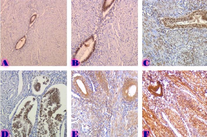
Endometrial Carcinoma is the most common gynaecological malignancy in the female population and is considered as incidentally the second gynaecological malignancy worldwide. Based on 2018 data more than 380 000 new cases were diagnosed worldwide and almost 90 000 of them had a lethal outcome. Interaction between cancer cells and their microenvironment regulates cancer progression in multiple types of cancer. It has great value in developing endometrial cancer and its progression respectively. There is no sufficient research data about the consequences and mechanisms which are participating in endometrial cancer progression and what determines its aggressive behaviour. Molecular signals derived from stromal cells and/or extracellular matrix plays a crucial role in malignancy. The cancer microenvironment is composed of cellular components and noncellular components (extracellular matrix)as well. Cancer cell invasion and metastasizing are some of the leading reasons why endometrial cancer is hardly sensitive to the treatment and has worse overall prognoses. Identification of Signaling pathways of the local microenvironment and peptides synthesized by stromal cells has a critical role in the modification of potentially significant biomarkers for endometrial cancer metastases and high-grade malignancy. In consideration of all of the mentioned microenvironment of endometrial cancer and its single components needs deeper examination while it has a critical value in understanding cancer aetiology, progression and its prognoses.
Downloads
Metrics
P. Morice, A. Leary, C. Creutzberg, N. Abu-Rustum, and E. Darai, “Endometrial cancer,” The Lancet, vol. 387, no. 10023, pp. 1094–1108, Mar. 2016, doi: 10.1016/S0140-6736(15)00130-0.
R. L. Siegel, K. D. Miller, and A. Jemal, “Cancer statistics, 2019,” CA: A Cancer Journal for Clinicians, vol. 69, no. 1, pp. 7–34, Jan. 2019, doi: 10.3322/CAAC.21551.
F. Bray, J. Ferlay, I. Soerjomataram, R. L. Siegel, L. A. Torre, and A. Jemal, “Global cancer statistics 2018: GLOBOCAN estimates of incidence and mortality worldwide for 36 cancers in 185 countries,” CA: A Cancer Journal for Clinicians, vol. 68, no. 6, pp. 394–424, Nov. 2018, doi: 10.3322/CAAC.21492.
G. D. Constantine, G. Kessler, S. Graham, and S. R. Goldstein, “Increased Incidence of Endometrial Cancer Following the Women’s Health Initiative: An Assessment of Risk Factors”, doi: 10.1089/jwh.2018.6956.
F. Guo, L. Levine, and A. Berenson, “Trends in the incidence of endometrial cancer among young women in the United States, 2001 to 2017.,” Journal of Clinical Oncology, vol. 39, no. 15_suppl, pp. 5578–5578, May 2021, doi: 10.1200/JCO.2021.39.15_SUPPL.5578.
“Rising Incidence of Endometrial Cancer Linked to Obesity in Younger Women - Cancer Therapy Advisor.” https://www.cancertherapyadvisor.com/home/news/conference-coverage/asco-2021/endometrial-cancer-obesity-younger-women-rising-incidence-risk/ (accessed Mar. 07, 2022).
F. J. A. Gujam, D. C. McMillan, Z. M. A. Mohammed, J. Edwards, and J. J. Going, “The relationship between tumour budding, the tumour microenvironment and survival in patients with invasive ductal breast cancer,” British Journal of Cancer, vol. 113, no. 7, pp. 1066–1074, Sep. 2015, doi: 10.1038/BJC.2015.287.
S. Rousset-Rouviere et al., “Endometrial Carcinoma: Immune Microenvironment and Emerging Treatments in Immuno-Oncology,” Biomedicines, vol. 9, no. 6, Jun. 2021, doi: 10.3390/BIOMEDICINES9060632.
S. Labiano, A. Palazon, and I. Melero, “Immune response regulation in the tumor microenvironment by hypoxia,” Seminars in Oncology, vol. 42, no. 3, pp. 378–386, Jun. 2015, doi: 10.1053/J.SEMINONCOL.2015.02.009.
D. Ribatti, G. Mangialardi, and A. Vacca, “Stephen Paget and the ‘seed and soil’ theory of metastatic dissemination,” Clinical and experimental medicine, vol. 6, no. 4, pp. 145–149, Dec. 2006, doi: 10.1007/S10238-006-0117-4.
I. Pastushenko and C. Blanpain, “EMT Transition States during Tumor Progression and Metastasis,” Trends in Cell Biology, vol. 29, no. 3, pp. 212–226, Mar. 2019, doi: 10.1016/J.TCB.2018.12.001.
S. Fulda, “Targeting Apoptosis Signaling Pathways for Anticancer Therapy,” Frontiers in Oncology, vol. 1, no. AUG, 2011, doi: 10.3389/FONC.2011.00023.
A. Markowska, M. Pawałowska, J. Lubin, and J. Markowska, “Signalling pathways in endometrial cancer,” Wspolczesna Onkologia, vol. 18, no. 3, pp. 143–148, 2014, doi: 10.5114/WO.2014.43154.
E. Coll-De La Rubia, E. Martinez-Garcia, G. Dittmar, A. Gil-Moreno, S. Cabrera, and E. Colas, “Clinical Medicine Prognostic Biomarkers in Endometrial Cancer: A Systematic Review and Meta-Analysis”, doi: 10.3390/jcm9061900.
H. Taghizadeh et al., “22P Hepatocyte growth factor (HGF) and c-Met expression in metastatic endometrial cancer,” Annals of Oncology, vol. 32, p. S1352, Oct. 2021, doi: 10.1016/J.ANNONC.2021.08.2018.
X. Shi, J. Wang, Y. Lei, C. Cong, D. Tan, and X. Zhou, “Research progress on the PI3K/AKT signaling pathway in gynecological cancer (Review),” Molecular Medicine Reports, vol. 19, no. 6, pp. 4529–4535, Jun. 2019, doi: 10.3892/MMR.2019.10121/HTML.
Q. Ping et al., “Cancer-associated fibroblasts: overview, progress, challenges, and directions,” Cancer Gene Therapy 2021 28:9, vol. 28, no. 9, pp. 984–999, Mar. 2021, doi: 10.1038/s41417-021-00318-4.
D. Zhao, X. ping Li, M. Gao, C. Zhao, J. liu Wang, and L. hui Wei, “Stromal cell-derived factor 1alpha stimulates human endometrial carcinoma cell growth through the activation of both extracellular signal-regulated kinase 1/2 and Akt,” Gynecologic oncology, vol. 103, no. 3, pp. 932–937, Dec. 2006, doi: 10.1016/J.YGYNO.2006.05.045.
X. Tang, “Tumor-associated macrophages as potential diagnostic and prognostic biomarkers in breast cancer,” Cancer Letters, vol. 332, no. 1, pp. 3–10, May 2013, doi: 10.1016/j.canlet.2013.01.024.
X. F. Jiang et al., “Tumor-associated macrophages correlate with progesterone receptor loss in endometrial endometrioid adenocarcinoma,” Journal of Obstetrics and Gynaecology Research, vol. 39, no. 4, pp. 855–863, Apr. 2013, doi: 10.1111/j.1447-0756.2012.02036.x.
J. Liu et al., “Association of tumour-associated macrophages with cancer cell EMT, invasion, and metastasis of Kazakh oesophageal squamous cell cancer,” Diagnostic Pathology, vol. 14, no. 1, Jun. 2019, doi: 10.1186/s13000-019-0834-0.
C. Casas-Arozamena and M. Abal, “Endometrial Tumour Microenvironment,” Advances in Experimental Medicine and Biology, vol. 1296, pp. 215–225, 2020, doi: 10.1007/978-3-030-59038-3_13.
X. Yang et al., “Increased expression of human macrophage metalloelastase (MMP-12) is associated with the invasion of endometrial adenocarcinoma,” Pathology Research and Practice, vol. 203, no. 7, pp. 499–505, Aug. 2007, doi: 10.1016/j.prp.2007.03.008.
G. D’Andrilli, A. Bovicelli, M. G. Paggi, and A. Giordano, “New insights in endometrial carcinogenesis,” Journal of Cellular Physiology, vol. 227, no. 7, pp. 2842–2846, Jul. 2012, doi: 10.1002/jcp.24016.
“Addressing activation of WNT beta-catenin pathway in diverse landscape of endometrial carcinogenesis - PubMed.” https://pubmed.ncbi.nlm.nih.gov/34956444/ (accessed Mar. 07, 2022).
D. Pradip, A. Jennifer, and D. Nandini, “Cancer-associated fibroblasts in conversation with tumor cells in endometrial cancers: A partner in crime,” International Journal of Molecular Sciences, vol. 22, no. 17, Sep. 2021, doi: 10.3390/ijms22179121.
C. M. Contreras et al., “Loss of Lkb1 provokes highly invasive endometrial adenocarcinomas,” Cancer Research, vol. 68, no. 3, pp. 759–766, Feb. 2008, doi: 10.1158/0008-5472.CAN-07-5014.
C. M. Contreras et al., “Lkb1 inactivation is sufficient to drive endometrial cancers that are aggressive yet highly responsive to mTOR inhibitor monotherapy,” DMM Disease Models and Mechanisms, vol. 3, no. 3–4, pp. 181–193, Mar. 2010, doi: 10.1242/dmm.004440.
C. G. Peña et al., “LKB1 loss promotes endometrial cancer progression via CCL2-dependent macrophage recruitment,” The Journal of clinical investigation, vol. 125, no. 11, pp. 4063–4076, Nov. 2015, doi: 10.1172/JCI82152.
H. Cheng et al., “A genetic mouse model of invasive endometrial cancer driven by concurrent loss of pten and Lkb1 is highly responsive to mTOR inhibition,” Cancer Research, vol. 74, no. 1, pp. 15–23, Jan. 2014, doi: 10.1158/0008-5472.CAN-13-0544.
K. van de Vijver, “Evalua&on of Tumor Infiltra&ng Lymphocytes (TILs) in Endometrial Carcinoma Guidelines for TILs assessment from the ‘Interna5onal Immuno-¬-Oncology Biomarker Working Group’”.
M. Tomšová, B. Melichar, I. Sedláková, and I. Šteiner, “Prognostic significance of CD3+ tumor-infiltrating lymphocytes in ovarian carcinoma.,” Gynecologic oncology, vol. 108, no. 2, pp. 415–420, Feb. 2008, doi: 10.1016/j.ygyno.2007.10.016.
W. Yamagami et al., “Immunofluorescence-detected infiltration of CD4+FOXP3+ regulatory T cells is relevant to the prognosis of patients with endometrial cancer.,” International journal of gynecological cancer : official journal of the International Gynecological Cancer Society, vol. 21, no. 9, pp. 1628–1634, Dec. 2011, doi: 10.1097/igc.0b013e31822c271f.
X. Yang et al., “Increased expression of human macrophage metalloelastase (MMP-12) is associated with the invasion of endometrial adenocarcinoma,” Pathology Research and Practice, vol. 203, no. 7, pp. 499–505, Aug. 2007, doi: 10.1016/j.prp.2007.03.008.
X. F. Jiang et al., “Tumor-associated macrophages correlate with progesterone receptor loss in endometrial endometrioid adenocarcinoma,” Journal of Obstetrics and Gynaecology Research, vol. 39, no. 4, pp. 855–863, Apr. 2013, doi: 10.1111/j.1447-0756.2012.02036.x.
E. C. Dun, K. Hanley, F. Wieser, S. Bohman, J. Yu, and R. N. Taylor, “Infiltration of tumor-associated macrophages is increased in the epithelial and stromal compartments of endometrial carcinomas,” International journal of gynecological pathology : official journal of the International Society of Gynecological Pathologists, vol. 32, no. 6, pp. 576–584, Nov. 2013, doi: 10.1097/PGP.0B013E318284E198.
Copyright (c) 2022 Paata Djordjoliani , Zaza Bokhua, George Burkadze

This work is licensed under a Creative Commons Attribution-NonCommercial-NoDerivatives 4.0 International License.


