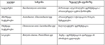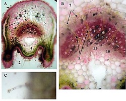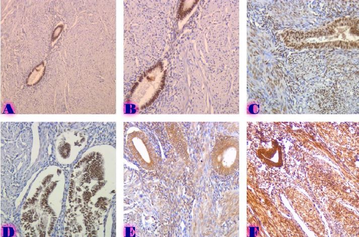
Evaluation of proliferative activity of pre-tumor and tumor processes of Barrett’s esophagus using AgNor technology
DOI:
https://doi.org/10.52340/gs.2023.05.02.07Keywords:
Barrett's esophagus, AgNOR, dysplasia, metaplasiaAbstract
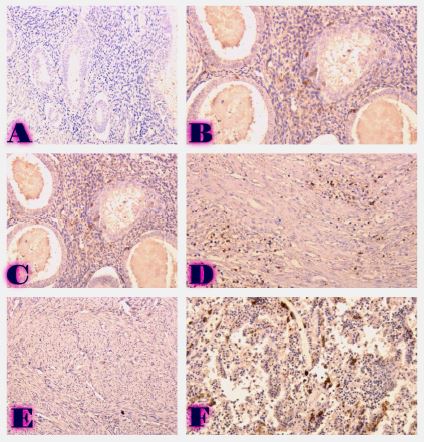
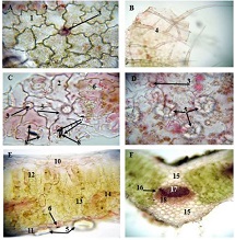
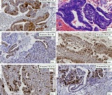
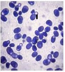
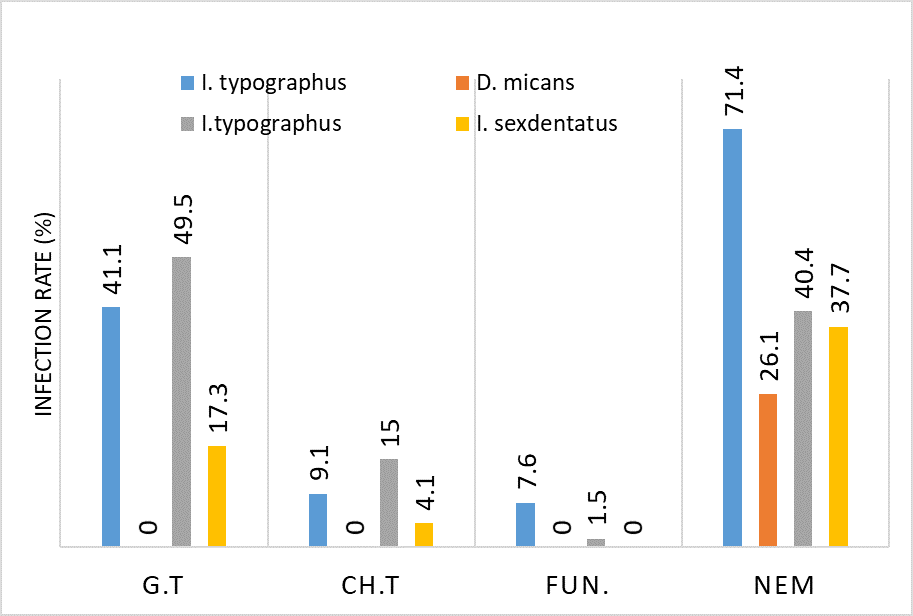
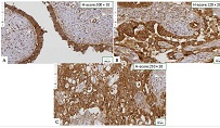
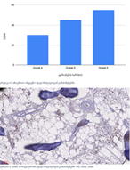
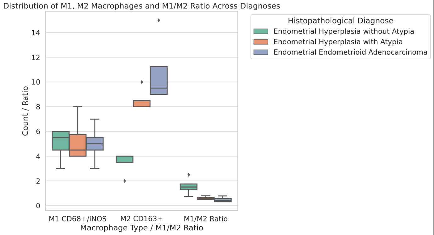
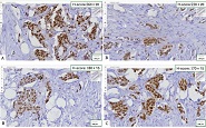
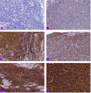
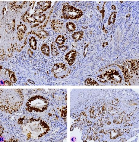
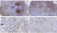
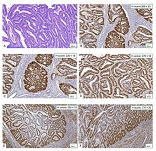
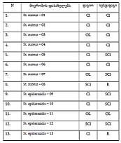
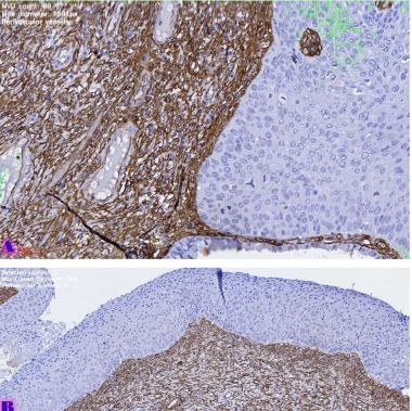
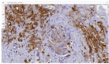
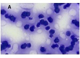
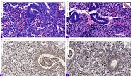
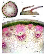
Esophageal carcinoma is the eighth most common malignancy and the sixth leading cause of cancer-related death worldwide. Adenocarcinomas account for the majority of esophageal carcinomas in the US. The incidence of esophageal squamous cell carcinomas is decreasing every year, while the number of adenocarcinomas has not changed over the last three to four decades. It has been established that there is a strong correlation between proliferative activity and a poor tumor prognosis, so the interest in clinical trials of proliferative potential on various markers is still relevant and increasing every year. Within the framework of our research, a cohort retrograde study was carried out, for which the archival material of the teaching-scientific and diagnostic laboratory of Tbilisi State Medical University for the years 2019-2022 was used. Proliferative activity was determined by AgNOR technology in 35 cases evaluated in the following histological entity: Barrett's esophagus with enteric metaplasia; Barrett's esophagus with foveolar metaplasia; Barrett's esophagus with low-grade dysplasia of the epithelium; Barrett's esophagus with high-grade dysplasia of the epithelium; Based on the results of our study, AgNOR technology can be used to evaluate proliferative activity in Barrett's esophagus.
Downloads
References
Siegel, R. L., Miller, K. D. & Jemal, A. Cancer statistics, 2020. CA Cancer J Clin 70, 7–30 (2020).
Sung, H. et al. Global Cancer Statistics 2020: GLOBOCAN Estimates of Incidence and Mortality Worldwide for 36 Cancers in 185 Countries. CA Cancer J Clin 71, 209–249 (2021).
Wani, S. et al. Risk factors for progression of low-grade dysplasia in patients with Barrett’s esophagus. Gastroenterology 141, (2011).
Song, Y. et al. Identification of Metastasis-Associated Biomarkers in Synovial Sarcoma Using Bioinformatics Analysis. Front Genet 11, 530892 (2020).
Kim, C. W. et al. Immunohistochemical expression of the p53 and Ki-67 proteins in Barrett’s esophagus in Korea. Korean J Gastroenterol 46, 189–195 (2005).
MESHVELIANI, P., DIDAVA, G., TOMADZE, G. & BURKADZE, G. CRITICAL REVIEW: BARRETS OESOPHAGUS – METAPLASIA – DYSPLASIA – MALIGNANT TRANSFORMAYION PHENOTYPICAL CHARACTERISTICS AND PROGRESSION MARKERS. EXPERIMENTAL AND CLINICAL MEDICINE GEORGIA (2022) doi:10.52340/JECM.2022.718.
Volkweis, B. S. et al. Ki-67 Antigen Overexpression Is Associated with the Metaplasia-Adenocarcinoma Sequence in Barrett’s Esophagus. Gastroenterol Res Pract 2012, (2012).
Szachnowicz, S. et al. Origin of adenocarcinoma in Barrett’s esophagus: p53 and Ki67 expression and histopathologic background. Clinics (Sao Paulo) 60, 103–112 (2005).
Goodarzi, M. et al. Anti-phosphorylated histone H3 expression in Barrett’s esophagus, low-grade dysplasia, high-grade dysplasia, and adenocarcinoma. Modern Pathology 22, 1612–1621 (2009).
Jankowski, J. A. et al. Molecular evolution of the metaplasia-dysplasia-adenocarcinoma sequence in the esophagus. American Journal of Pathology 154, 965–973 (1999).
Shaheen, N. J. Advances in Barrett’s esophagus and esophageal adenocarcinoma. Gastroenterology 128, 1554–1566 (2005).
Karamchandani, D. M. et al. Increasing diagnostic accuracy to grade dysplasia in Barrett’s esophagus using an immunohistochemical panel for CDX2, p120ctn, c-Myc and Jagged1. Diagn Pathol 11, (2016).
Metreveli, B., Gagua, D., Burkadze, G. & Kepuladze, S. PROLIFERATIVE CHARACTERISTICS OF EUTOPIC AND ECTOPIC ENDOMETRIUM IN ADENOMYOSIS USING AgNOR TECHNOLOGY. GEORGIAN SCIENTISTS 5, 59–71 (2023).
Tavdgiridze, N., Tevdorashvili, G., Kepuladze, S. & Burkadze, G. ASSESSMENT OF PROLIFERATIVE ACTIVITY OF IMMATURE OVARIAN TERATOMAS USING AgNOR TECHNOLOGY. GEORGIAN SCIENTISTS 5, 233–248 (2023).
Murgod, S., Channabasaviah, G. H., Shivamurthy, D. M., Ashok, L. & Krishnappa, S. J. Prognostic potential of AgNORs in oral submucous fibrosis. J Int Soc Prev Community Dent 6, 167–75 (2016).
Nishie, H., Hirooka, Y. & Kaibara, N. Argyrophilic Nucleolar Organizer Regions of Breast Tumors: Assessment of Aspirated Cytological Materials. Breast Cancer 3, 189–204 (1996).
Chiu, K. Y., Loke, S. L. & Wong, K. K. Improved silver technique for showing nucleolar organiser regions in paraffin wax sections. J Clin Pathol 42, 992–994 (1989).
Downloads
Published
How to Cite
Issue
Section
License
Copyright (c) 2023 GEORGIAN SCIENTISTS

This work is licensed under a Creative Commons Attribution-NonCommercial-NoDerivatives 4.0 International License.