The histopathological and phenotypic characteristics of keloid and hypertrophic scar during wound closure with compressive and non-compressive sutures
Downloads
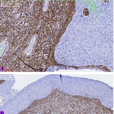
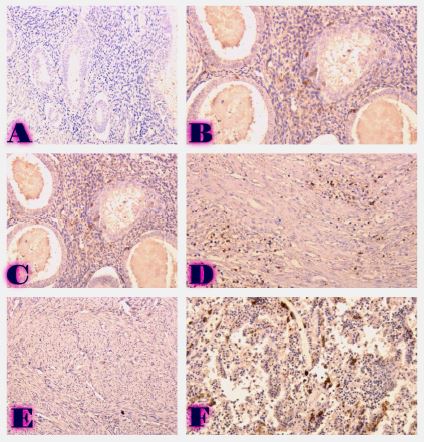
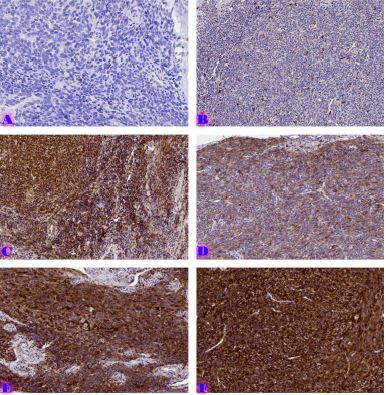
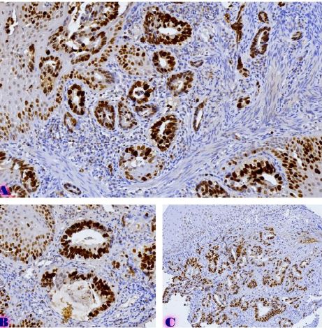
Keloids and hypertrophic scars are fibroproliferative disorders caused by abnormal wound healing. They extend beyond the borders of the primary wound, do not regress spontaneously, and tend to recur after excision. Every year in developed countries, about 100 million people face problems related to scars. Hypertrophied scars and keloids have a significant impact on the patient's quality of life, physical condition and psychological health. Compression sutures are traditionally used for wound closure in clinical practice. Compression sutures mean regular sutures, which are used to close the wound with a knotted suture, which causes tissue compression. Non-compressive sutures are sutures of ordinary composition treated with a unique technology, which have a tufted structure, and at the expense of these tufts, they close the wound without forming knots. In case of their use, the histopathological and phenotypic features of the development of keloids and skin scars compared to compressive threads require more research. In histopathological practice, the histopathological indicators that will allow us to determine the high and low risk groups for the development of keloids and hypertrophic scars are still not well studied; Which may have a great clinical value because it allowed us to identify the high and low risk of possible development of keloid based on Punch-biopsy. The complex mechanisms of keloid formation are still unknown and probably depend on the body's ability to respond to damage on the one hand and environmental factors, including the use of compression threads, on the other hand.
Downloads
T. Zhang et al., “Current potential therapeutic strategies targeting the TGF-β/Smad signaling pathway to attenuate keloid and hypertrophic scar formation,” Biomedicine and Pharmacotherapy, vol. 129. Elsevier Masson s.r.l., Sep. 01, 2020. doi: 10.1016/j.biopha.2020.110287.
Z. C. Wang et al., “The Roles of Inflammation in Keloid and Hypertrophic Scars,” Frontiers in Immunology, vol. 11. Frontiers Media S.A., Dec. 04, 2020. doi: 10.3389/fimmu.2020.603187.
A. Shaheen, “Comprehensive Review of Keloid Formation,” Journal of Clinical Research in Dermatology, vol. 4, no. 5, pp. 1–18, Oct. 2017, doi: 10.15226/2378-1726/4/5/00168.
R. Ogawa, “Keloid and hypertrophic scars are the result of chronic inflammation in the reticular dermis,” International Journal of Molecular Sciences, vol. 18, no. 3. MDPI AG, Mar. 10, 2017. doi: 10.3390/ijms18030606.
R. Ogawa and S. Akaishi, “Endothelial dysfunction may play a key role in keloid and hypertrophic scar pathogenesis – Keloids and hypertrophic scars may be vascular disorders,” Med Hypotheses, vol. 96, pp. 51–60, Nov. 2016, doi: 10.1016/j.mehy.2016.09.024.
Walraven M., “Cellular and molecular mechanisms involved in scarless wound healing in the fetal skin.,” Amsterdam: VU university medical center, 2016.
W. Lv et al., “Epigenetic modification mechanisms involved in keloid: current status and prospect,” Clinical Epigenetics, vol. 12, no. 1. BioMed Central Ltd, Dec. 01, 2020. doi: 10.1186/s13148-020-00981-8.
T. Unahabhokha, A. Sucontphunt, U. Nimmannit, P. Chanvorachote, N. Yongsanguanchai, and V. Pongrakhananon, “Molecular signalings in keloid disease and current therapeutic approaches from natural based compounds,” Pharmaceutical Biology, vol. 53, no. 3. Informa Healthcare, pp. 457–463, Mar. 01, 2015. doi: 10.3109/13880209.2014.918157.
S. Tan, N. Khumalo, and A. Bayat, “Understanding keloid pathobiology from a quasi-neoplastic perspective: Less of a scar and more of a chronic inflammatory disease with cancer-like tendencies,” Frontiers in Immunology, vol. 10. Frontiers Media S.A., 2019. doi: 10.3389/fimmu.2019.01810.
A. S. Vincent, T. T. Phan, A. Mukhopadhyay, H. Y. Lim, B. Halliwell, and K. P. Wong, “Human skin keloid fibroblasts display bioenergetics of cancer cells,” Journal of Investigative Dermatology, vol. 128, no. 3, pp. 702–709, 2008, doi: 10.1038/sj.jid.5701107.
T. Zhang et al., “Current potential therapeutic strategies targeting the TGF-β/Smad signaling pathway to attenuate keloid and hypertrophic scar formation,” Biomedicine and Pharmacotherapy, vol. 129. Elsevier Masson s.r.l., Sep. 01, 2020. doi: 10.1016/j.biopha.2020.110287.
D. Son and A. Harijan, “Overview of surgical scar prevention and management,” Journal of Korean Medical Science, vol. 29, no. 6. Korean Academy of Medical Science, pp. 751–757, 2014. doi: 10.3346/jkms.2014.29.6.751.
Harn HI, Ogawa R, Hsu CK, Hughes MW, Tang MJ, and Chuong CM, “The tension biology of wound healing.,” Exp Dermatol., 2017.
L. Téot, T. A. Mustoe, E. Middelkoop, and G. G. Gauglitz, “State of the Art Management and Emerging Technologies Textbook on Scar Management 123.”
Arima J, Dohi T, Kuribayashi S, Akaishi S, and Ogawa R, “Z-plasty and postoperative radiation therapy for anterior chest wall, keloids: an analysis of 141 patients.,” Plast Reconstr Surg Glob, 2019.

This work is licensed under a Creative Commons Attribution-NonCommercial-NoDerivatives 4.0 International License.


