Assessment of FOXP3+ Expression in Endometrial Precancerous and Neoplastic Lesions: A Comparative Study of Immune Microenvironment Dynamics
Загрузки
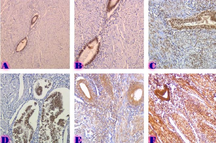
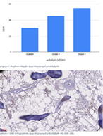
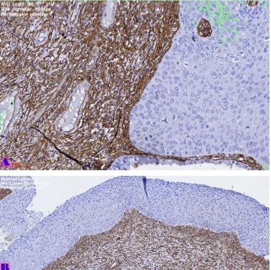
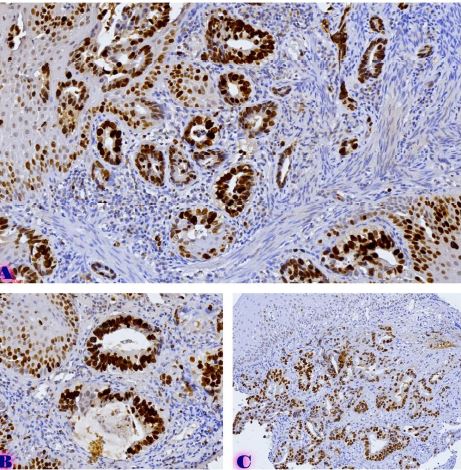
Background: Endometrial carcinoma is a leading cause of cancer-related mortality in women, with distinct subtypes such as endometrioid and serous carcinoma exhibiting varying clinical behaviours. Regulatory T cells (Tregs), marked by FOXP3 expression, play a crucial role in immune modulation within the tumour microenvironment. Programmed cell death ligand 1 (PDL1) is another key immune checkpoint molecule associated with immune evasion in various malignancies. This study investigates the expression of FOXP3 and PDL1 in benign and malignant endometrial lesions, comparing endometrial hyperplasias, endometrioid adenocarcinoma, and serous carcinoma. Methods: 150 cases were analysed, including 80 benign hyperplasias (with and without atypia) and 70 malignant lesions (endometrioid and serous carcinoma). FOXP3 and PDL1 expression levels were assessed using immunohistochemistry, and statistical analyses were performed to evaluate the correlation between these markers and clinicopathological features such as age, tumour stage, and histological subtype. Results: FOXP3 expression was significantly higher in malignant lesions (mean 22-30%) compared to benign hyperplasias (mean 5%). Serous carcinoma exhibited the highest FOXP3 expression (30%), followed by endometrioid carcinoma (22%). PDL1 expression was also significantly higher in malignant tumours, with serous carcinoma showing a mean expression of 12% compared to 6% in endometrioid carcinoma. A positive correlation between FOXP3 and PDL1 expression was observed (Spearman’s correlation coefficient = 0.75, p<0.001). Additionally, higher FOXP3 and PDL1 expressions were observed in advanced-stage tumours (pT2) compared to early-stage tumours (pT1). Conclusion: The study demonstrates that FOXP3-positive Tregs and PDL1 are significantly upregulated in malignant endometrial tumours, particularly in serous carcinoma, associated with a more aggressive clinical course. The positive correlation between FOXP3 and PDL1 expression suggests a potential mechanism of immune evasion and supports immune checkpoint inhibition as a therapeutic strategy in endometrial carcinoma.
Скачивания
A. S. Uduwela, M. A. K. Perera, L. Aiqing, and I. S. Fraser, “Endometrial-myometrial interface: Relationship to adenomyosis and changes in pregnancy,” Obstet Gynecol Surv, vol. 55, no. 6, pp. 390–400, Jun. 2000, doi: 10.1097/00006254-200006000-00025.
P. Morice, A. Leary, C. Creutzberg, N. Abu-Rustum, and E. Darai, “Endometrial cancer,” The Lancet, vol. 387, no. 10023, pp. 1094–1108, Mar. 2016, doi: 10.1016/S0140-6736(15)00130-0.
C. Casas-Arozamena and M. Abal, “Endometrial Tumour Microenvironment,” Adv Exp Med Biol, vol. 1296, pp. 215–225, 2020, doi: 10.1007/978-3-030-59038-3_13.
G. D’Andrilli, A. Bovicelli, M. G. Paggi, and A. Giordano, “New insights in endometrial carcinogenesis,” J Cell Physiol, vol. 227, no. 7, pp. 2842–2846, Jul. 2012, doi: 10.1002/jcp.24016.
A. Markowska, M. Pawałowska, J. Lubin, and J. Markowska, “Signalling pathways in endometrial cancer,” Wspolczesna Onkologia, vol. 18, no. 3, pp. 143–148, 2014, doi: 10.5114/WO.2014.43154.
P. Zhorzholiani, Z. Bokhua, S. Kepuladze, and G. Burkadze, “The Role of M1 and M2 Macrophages in the Progression of Endometrial Hyperplasia to Endometrioid Adenocarcinoma,” ქართველი მეცნიერები, vol. 7, no. 1, pp. 24–37, Jan. 2025, doi: 10.52340/gs.2025.07.01.04.
M. Arafat Hossain, “A comprehensive review of immune checkpoint inhibitors for cancer treatment,” Int Immunopharmacol, vol. 143, no. Pt 2, Dec. 2024, doi: 10.1016/J.INTIMP.2024.113365.
Y. Wang and B. P. Zhou, “Epithelial-mesenchymal transition in breast cancer progression and metastasis,” Chin J Cancer, vol. 30, no. 9, p. 603, 2011, doi: 10.5732/CJC.011.10226.
L. Uroog, A. H. Rahmani, M. A. Alsahli, S. A. Almatroodi, R. A. Wani, and M. Moshahid Alam Rizvi, “Genetic Profile of FOXO3 Single-Nucleotide Polymorphism in Colorectal Cancer Patients,” Oncology (Switzerland), vol. 102, no. 4, pp. 299–309, Oct. 2023, doi: 10.1159/000533729/867502/GENETIC-PROFILE-OF-FOXO3-SINGLE-NUCLEOTIDE.
S. Sakaguchi, T. Yamaguchi, T. Nomura, and M. Ono, “Regulatory T cells and immune tolerance,” Cell, vol. 133, no. 5, pp. 775–787, May 2008, doi: 10.1016/J.CELL.2008.05.009.
D. Tan, Y. Fu, W. Tong, and F. Li, “Prognostic significance of lymphocyte to monocyte ratio in colorectal cancer: A meta-analysis,” International Journal of Surgery, vol. 55, pp. 128–138, Jul. 2018, doi: 10.1016/J.IJSU.2018.05.030.
S. L. Topalian et al., “Safety, activity, and immune correlates of anti-PD-1 antibody in cancer,” N Engl J Med, vol. 366, no. 26, pp. 2443–2454, Jun. 2012, doi: 10.1056/NEJMOA1200690.
R. Saleh and E. Elkord, “FoxP3+ T regulatory cells in cancer: Prognostic biomarkers and therapeutic targets,” Cancer Lett, vol. 490, pp. 174–185, Oct. 2020, doi: 10.1016/J.CANLET.2020.07.022.
Y. Ohue and H. Nishikawa, “Regulatory T (Treg) cells in cancer: Can Treg cells be a new therapeutic target?,” Cancer Sci, vol. 110, no. 7, pp. 2080–2089, Jul. 2019, doi: 10.1111/CAS.14069.
Copyright (c) 2025 Georgian Scientists

Это произведение доступно по лицензии Creative Commons «Attribution-NonCommercial-NoDerivatives» («Атрибуция — Некоммерческое использование — Без производных произведений») 4.0 Всемирная.


