Proliferative characteristics of eutopic and ectopic endometrium in adenomyosis using AgNOR technology

Downloads


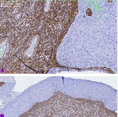
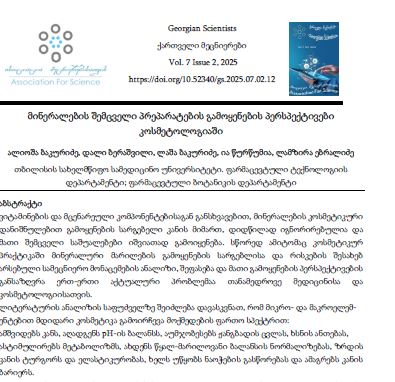
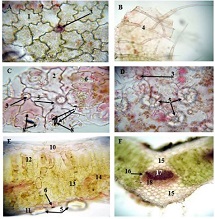
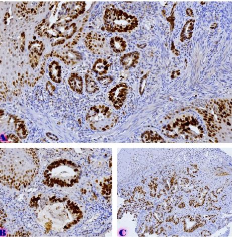
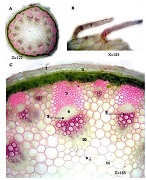
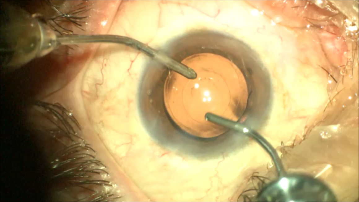
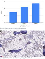
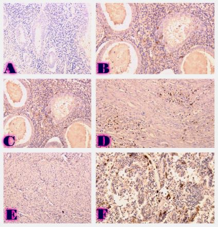
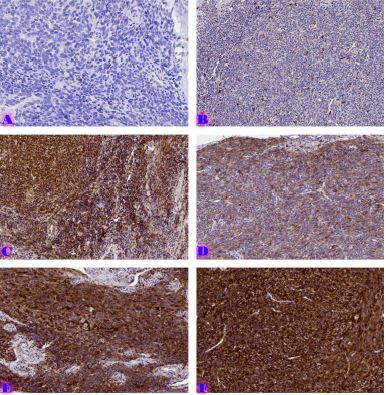
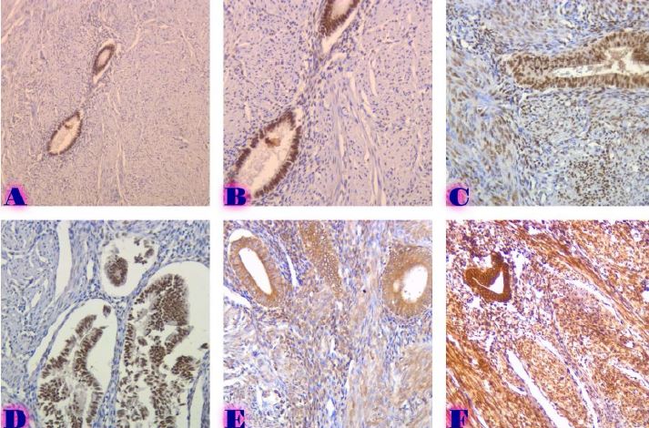
Adenomyosis is a benign lesion of the uterus, which is characterized by the presence and pathological growth of endometrial glands and stroma in the myometrium. During adenomyosis, endometrial tissue abnormally grown in the myometrium is called ectopic endometrium, and endometrial tissue in the uterine cavity is called eutopic endometrium. Women with adenomyosis may experience abnormal uterine bleeding, dysmenorrhea, dyspareunia, and infertility, although, in one-third of women, the lesion is asymptomatic. Adenomyosis is the most frequently diagnosed pathology after hysterectomy in perimenopausal women due to heavy bleeding or pain. AgNOR is used as an aid for evaluating the proliferative activity of cells. Certain types of studies have demonstrated their effectiveness in differentiating various benign or malignant processes, as well as in determining the histological grade of intraepithelial neoplastic processes. Within the framework of this study, the proliferative activity of the eutopic and ectopic endometrium, as well as their stroma and normal myometrium, was evaluated on archival blocks fixed in formalin and embedded in paraffin (FFPE), diagnosed in cases of adenomyosis, using AgNOR technology. In the studied system, eutopic endometrium - basal endometrium - in both the glandular and stromal components of ectopic endometrium, the highest proliferative activity is manifested in ectopic endometrium, which is why it can probably represent a risk for the development of neoplasms.
Downloads
Antero MF, Ayhan A, Segars J, Shih IM. Pathology and Pathogenesis of Adenomyosis. Semin Reprod Med. 2020 May 1;38(2–3):108–18.
Devlieger R, D’Hooghe T, Timmerman D. Uterine adenomyosis in the infertility clinic. Hum Reprod Update. 2003 Mar;9(2):139–47.
Yamaguchi M, Yoshihara K, Suda K, Nakaoka H, Yachida N, Ueda H, et al. Three-dimensional understanding of the morphological complexity of the human uterine endometrium. iScience. 2021 Apr 23;24(4).
Chapron C, Marcellin L, Borghese B, Santulli P. Rethinking mechanisms, diagnosis and management of endometriosis. Nat Rev Endocrinol. 2019 Nov 1;15(11):666–82.
García-Solares J, Donnez J, Donnez O, Dolmans MM. Pathogenesis of uterine adenomyosis: invagination or metaplasia? Fertil Steril. 2018 Mar 1;109(3):371–9.
Camboni A, Marbaix E. Ectopic endometrium: The pathologist’s perspective. Int J Mol Sci. 2021 Oct 1; 22(20).
Munro MG. Classification and Reporting Systems for Adenomyosis. J Minim Invasive Gynecol. 2020 Feb 1; 27(2):296–308.
Habiba M, Gordts S, Bazot M, Brosens I, Benagiano G. Exploring the challenges for a new classification of adenomyosis. Reprod Biomed Online. 2020 Apr 1;40(4):569–81.
Goldblum JR, Clement PB, Hart WR. Adenomyosis with sparse glands: A potential mimic of low-grade endometrial stromal sarcoma. Am J Clin Pathol. 1995;103(2):218–23.
Hirschowitz L, Mayall FG, Ganesan R, McCluggage WG. Intravascular adenomyomatosis expanding the morphologic spectrum of intravascular leiomyomatosis. American Journal of Surgical Pathology. 2013 Sep;37(9):1395–400.
Uduwela AS, Perera MAK, Aiqing L, Fraser IS. Endometrial-myometrial interface: Relationship to adenomyosis and changes in pregnancy. Obstet Gynecol Surv. 2000 Jun;55(6):390–400.
KY Chiu SLKW. Improved silver technique for showing nucleolar organiser regions in paraffin wax sections. J Clin Pathol. 1989;42:992–4.
Boquist L. Nucleolar organizer regions in normal, hyperplastic and neoplastic parathyroid glands. Virchows Archiv [A] Pathol Anat. 1990;417:237–41.
Caldeira PC, Aguiar MCF, Mesquita RA, do Carmo MAV. Oral leukoplakias with different degrees of dysplasia: Comparative study of hMLH1, p53, and AgNOR. Journal of Oral Pathology and Medicine. 2011 Apr;40(4):305–11.
Ploton D, Menager M, Jeannesson P, Himber G, Pigeon F, Adnet JJ. Improvement in the staining and in the visualization of the argyrophilic proteins of the nucleolar organizer region at the optical level. Histochem J. 1986 Jan;18(1):5–14.
Li Q, Hacker GW, Danscher G, Sonnleitner-Wittauer U, Grimelius L. Argyrophilic nucleolar organizer regions - A revised version of the Ag-NOR-staining technique. Histochem Cell Biol [Internet]. 1995 Aug [cited 2023 Jan 22];104(2):145–50. Available from: https://link.springer.com/article/10.1007/BF01451573
Ferreira SJ, Machado MÂN, de Lima AAS, Johann ACBR, Grégio AMT, Azevedo-Alanis LR. Identification of AgNORs and cytopathological changes in oral lichen planus lesions. Acta Histochem. 2017 Jan 1;119(1):32–8.
Harsh A, Tondon R, Harsh HK, Professor E. Utility of AgNOR Count in Non-Neoplastic and Neoplastic Lesions of the Uterine Cervix. Original Research Article [Internet]. 2018 [cited 2022 Dec 30];198(1):198–201. Available from: www.ijmrp.com
Rüschoff J, Plate K, Bittinger A, Thomas C. Nucleolar Organizer Regions (NORs): Basic Concepts and Practical Application in Tumor Pathology. Pathol Res Pract. 1989;185(6):878–85.

This work is licensed under a Creative Commons Attribution-NonCommercial-NoDerivatives 4.0 International License.