Study of inflammatory-fibrotic index of keloids and hypertrophic scars and features of proliferative activity of fibroblasts
Downloads
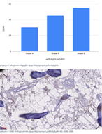
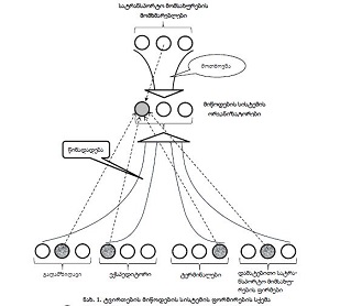
Wound healing is a physiological, complex, and well-regulated process involving a combination of cell migration, inflammation, innervation, and angiogenesis processes. Keloids (KS) and hypertrophic scars (HS) are formed as a result of damage to the epidermis and dermis. Although their exact pathogenesis is still not fully known, it is widely hypothesized that dysregulation of the molecular mechanisms involved in wound healing may lead to the formation of skin scars. Any deviation from the normal wound healing response can lead to excessive collagen accumulation and improper remodeling of the extracellular matrix (ECM), leading to the formation of a keloid or hypertrophic scar. The aim of our study was to study the characteristics of the distribution of inflammatory indices in dermatofibroma, keloid and hypertrophic scars, as well as to determine its relationship with the proliferative activity detected by AgNOR technology.
Downloads
Gauglitz GG, Korting HC, Pavicic T, Ruzicka T, Jeschke MG. Hypertrophic Scarring and Keloids: Pathomechanisms and Current and Emerging Treatment Strategies. Molecular Medicine [Internet]. 2011 Jan [cited 2024 Feb 17];17(1–2):113. Available from: /pmc/articles/PMC3022978/
Shaheen A. Comprehensive Review of Keloid Formation. Journal of Clinical Research in Dermatology. 2017 Oct 12;4(5):1–18.
Tan S, Khumalo N, Bayat A. Understanding Keloid Pathobiology From a Quasi-Neoplastic Perspective: Less of a Scar and More of a Chronic Inflammatory Disease With Cancer-Like Tendencies. Front Immunol. 2019 Aug 7;10.
Huang C, Murphy GF, Akaishi S, Ogawa R. Keloids and Hypertrophic Scars: Update and Future Directions. Plast Reconstr Surg Glob Open [Internet]. 2013 Jul 2 [cited 2024 Feb 17];1(4). Available from: /pmc/articles/PMC4173836/
Lee JYY, Yang CC, Chao SC, Wong TW. Histopathological differential diagnosis of keloid and hypertrophic scar. Am J Dermatopathol [Internet]. 2004 Oct [cited 2024 Feb 17];26(5):379–84. Available from: https://pubmed.ncbi.nlm.nih.gov/15365369/
Limandjaja GC, Niessen FB, Scheper RJ, Gibbs S. Hypertrophic scars and keloids: Overview of the evidence and practical guide for differentiating between these abnormal scars. Exp Dermatol. 2021 Jan 1;30(1):146–61.
Ogawa R. Keloid and Hypertrophic Scars Are the Result of Chronic Inflammation in the Reticular Dermis. Int J Mol Sci [Internet]. 2017 Mar 10 [cited 2024 Feb 17];18(3). Available from: https://pubmed.ncbi.nlm.nih.gov/28287424/
Sarrazy V, Billet F, Micallef L, Coulomb B, Desmoulière A. Mechanisms of pathological scarring: role of myofibroblasts and current developments. Wound Repair Regen [Internet]. 2011 [cited 2024 Feb 17];19 Suppl 1(SUPPL. 1):s10–5. Available from: https://pubmed.ncbi.nlm.nih.gov/21793960/
Ogawa R. Keloid and Hypertrophic Scars Are the Result of Chronic Inflammation in the Reticular Dermis. Int J Mol Sci [Internet]. 2017 Mar 10 [cited 2024 Feb 17];18(3). Available from: https://pubmed.ncbi.nlm.nih.gov/28287424/
Huang C, Akaishi S, Hyakusoku H, Ogawa R. Are keloid and hypertrophic scar different forms of the same disorder? A fibroproliferative skin disorder hypothesis based on keloid findings. Int Wound J [Internet]. 2014 Oct 1 [cited 2024 Feb 17];11(5):517–22. Available from: https://pubmed.ncbi.nlm.nih.gov/23173565/
Adamashvili, N., Beriashvili, R., Tevzadze, N., Kepuladze, S., & Burkadze, G. (2024). Features of the distribution of acute and chronic inflammatory index in cervical intraepithelial neoplasia of different degrees and its relationship with proliferative activity detected by AgNOR technology. Georgian Scientists, 6(1), 45–57. https://doi.org/10.52340/gs.2024.06.01.08
Meshveliani, P., Didava, G., Tomadze, G., Kepuladze, S., & Burkadze, G. (2023). Evaluation of proliferative activity of pre-tumor and tumor processes of Barrett’s esophagus using AgNor technology. Georgian Scientists, 5(2), 49–62. https://doi.org/10.52340/gs.2023.05.02.07
Kveliashvili, T., Didava, G., Tevzadze, N., Kepuladze, S., & Burkadze, G. (2023). Peculiarities of the proliferative activity of the gallbladder mucosa in precancerous and cancerous pathologies detected by AgNOR technology. Georgian Scientists, 5(4), 342–352. https://doi.org/10.52340/gs.2023.05.04.31
Turashvili, T., Tevdorashvili, G., Burkadze, G., & Kepuladze, S. (2023). Evaluation of proliferative activity of endometrial metaplasias by AgNor technology. Georgian Scientists, 5(3), 10–20. https://doi.org/10.52340/2023.05.03.02
Svanadze T, Kepuladze S, Tevzadze N, Burkadze G. Assessment of proliferative activity of different types of squamous cell metaplasia of the cervix using agnor technology. Georgian Scientists, 5(2), 275–287. https://doi.org/10.52340/gs.2023.05.02.35
Arveladze, G., Beriashvili, R., Kepuladze, S., & Burkadze, G. (2023). Evaluation of proliferative activity of different subtypes of basal cell carcinomas by AgNOR technology. Georgian Scientists, 5(4), 206–218. https://doi.org/10.52340/gs.2023.05.04.19
Tavdgiridze N, Tevdorashvili G, Kepuladze S, Burkadze G. Assessment of proliferative activity of immature ovarian teratomas using AgNOR technology. Georgian Scientists, 5(1), 233–248. https://doi.org/10.52340/gs.2023.05.01.20
Metreveli B, Gagua D, Burkadze G, Kepuladze S. Proliferative characteristics of eutopic and ectopic endometrium in adenomyosis using AgNOR technology. Georgian Scientists, 5(1), 59–71. https://doi.org/10.52340/gs.2023.05.01.04
Copyright (c) 2024 Georgian Scientists

This work is licensed under a Creative Commons Attribution-NonCommercial-NoDerivatives 4.0 International License.


