Atypical hyperplasia of the endocervix - problematic issues related to molecular characteristics of adenocarcinoma, risk of progression and metastasis
Downloads
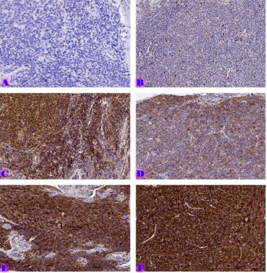
Cervical cancer is considered a multifactorial disease that includes socio-economic, cultural, immunological and epigenetic factors. Tumour precursor lesions, which are the basis for the development of endocervical tumours, have been studied. However, the molecular basis of this process is still under investigation. Unlike squamous cell carcinomas, cervical adenocarcinomas comprise a heterogeneous group of tumours that are not universally associated with HPV infection. These tumours show different morphology, aetiology, and prognosis. It is important to study and deeply analyse the pathogenesis, molecular, and immunohistochemical characteristics of endocervix adenocarcinomas, and precursor lesions associated with them. Studies have shown that tumour stem cells significantly affect the tumour's size, the progression rate, and the regression degree after treatment. Tumour proliferative activity, apoptotic status, and tumour stem cells are thought to be associated with poor prognosis and drug resistance in cervical cancer. All of the above confirms the existence of a complex regulatory mechanism underlying the development and progression of cervical cancer. Therefore, detecting biomarkers for each stage of the disease is necessary to improve the current knowledge and develop methods for early diagnosis and therapy in the future.
Downloads
F. Bray, J. Ferlay, I. Soerjomataram, R. L. Siegel, L. A. Torre, and A. Jemal, “Global cancer statistics 2018: GLOBOCAN estimates of incidence and mortality worldwide for 36 cancers in 185 countries.,” CA Cancer J Clin, vol. 68, no. 6, pp. 394–424, Nov. 2018, doi: 10.3322/caac.21492.
“https://www.ncdc.ge/#/pages/file/ea1784b5-d3d0-4dd9-b29f-1369f5d6bbec”.
A. D. Campos-Parra et al., “Molecular Differences between Squamous Cell Carcinoma and Adenocarcinoma Cervical Cancer Subtypes: Potential Prognostic Biomarkers,” Current Oncology, vol. 29, no. 7, pp. 4689–4702, Jul. 2022, doi: 10.3390/curroncol29070372.
Y. Zhou, W. Wang, K. Hu, and F. Zhang, “Comparison of Outcomes and Prognostic Factors Between Early-Stage Cervical Adenocarcinoma and Adenosquamous Carcinoma Patients After Radical Surgery and Postoperative Adjuvant Radiotherapy,” Cancer Manag Res, vol. Volume 13, pp. 7597–7605, Oct. 2021, doi: 10.2147/CMAR.S329614.
M. Wang, B. Yuan, Z. Zhou, and W. Han, “Clinicopathological characteristics and prognostic factors of cervical adenocarcinoma,” Sci Rep, vol. 11, no. 1, p. 7506, Apr. 2021, doi: 10.1038/s41598-021-86786-y.
Carcangiu M, Herrington C, Young R, and Kurman R, “WHO Classification of Tumours of Female Reproductive Organs.,” France: IARC Press, 2014.
S. Stolnicu et al., “International Endocervical Adenocarcinoma Criteria and Classification (IECC),” American Journal of Surgical Pathology, vol. 42, no. 2, pp. 214–226, Feb. 2018, doi: 10.1097/PAS.0000000000000986.
A. Hodgson and K. J. Park, “Cervical Adenocarcinomas: A Heterogeneous Group of Tumors With Variable Etiologies and Clinical Outcomes,” Arch Pathol Lab Med, vol. 143, no. 1, pp. 34–46, Jan. 2019, doi: 10.5858/arpa.2018-0259-RA.
J. Schwock et al., “Stratified Mucin-Producing Intraepithelial Lesion of the Cervix: Subtle Features Not to Be Missed,” Acta Cytol, vol. 60, no. 3, pp. 225–231, 2016, doi: 10.1159/000447940.
S. Stolnicu et al., “International Endocervical Adenocarcinoma Criteria and Classification (IECC),” American Journal of Surgical Pathology, vol. 42, no. 2, pp. 214–226, Feb. 2018, doi: 10.1097/PAS.0000000000000986.
C. Carleton et al., “A Detailed Immunohistochemical Analysis of a Large Series of Cervical and Vaginal Gastric-type Adenocarcinomas,” American Journal of Surgical Pathology, vol. 40, no. 5, pp. 636–644, May 2016, doi: 10.1097/PAS.0000000000000578.
Howitt BE and Nucci MR, “Mesonephric proliferations of the female genital tract.,” Pathology (Phila), 2018.
Mirkovic J, Schoolmeester JK, Campbell F, Miron A, Nucci MR, and Howitt BE, “Cervical mesonephric hyperplasia lacks KRAS/NRAS mutations.,” Histopathology, 2017.
D. Huo, D. Anderson, J. R. Palmer, and A. L. Herbst, “Incidence rates and risks of diethylstilbestrol-related clear-cell adenocarcinoma of the vagina and cervix: Update after 40-year follow-up,” Gynecol Oncol, vol. 146, no. 3, pp. 566–571, Sep. 2017, doi: 10.1016/j.ygyno.2017.06.028.
C. J. R. Stewart, M. H. E. Koay, C. Leslie, N. Acott, and Y. C. Leung, “Cervical carcinomas with a micropapillary component: a clinicopathological study of eight cases,” Histopathology, vol. 72, no. 4, pp. 626–633, Mar. 2018, doi: 10.1111/his.13419.
S. Stolnicu et al., “International Endocervical Adenocarcinoma Criteria and Classification (IECC),” American Journal of Surgical Pathology, vol. 42, no. 2, pp. 214–226, Feb. 2018, doi: 10.1097/PAS.0000000000000986.
J. Schwock et al., “Stratified Mucin-Producing Intraepithelial Lesion of the Cervix: Subtle Features Not to Be Missed,” Acta Cytol, vol. 60, no. 3, pp. 225–231, 2016, doi: 10.1159/000447940.
D. S. Heller, L. Nguyen, and L. T. Goldsmith, “Association of Cervical Microglandular Hyperplasia With Exogenous Progestin Exposure.,” J Low Genit Tract Dis, vol. 20, no. 2, pp. 162–4, Apr. 2016, doi: 10.1097/LGT.0000000000000176.
A. Molero, A. Parra, I. Blanco, A. Ascensión, and P. Ortega, “Lobular Endocervical Glandular Hyperplasia, a mimicker and potential pitfall for HPV-independent well differentiated Gastric-type Endocervical Adenocarcinoma: Case report and literature review focusing on histology, immunophenotype, and molecular findings.,” SAGE Open Med Case Rep, vol. 11, p. 2050313X231186210, 2023, doi: 10.1177/2050313X231186210.
K. Ida et al., “Whole-exome sequencing of lobular endocervical glandular hyperplasia.,” Oncol Lett, vol. 18, no. 3, pp. 2592–2597, Sep. 2019, doi: 10.3892/ol.2019.10549.
S. F. Roy, J. Wong, M. Latour, B. Reichetzer, and K. Rahimi, “Ductal-type mesonephric duct/remnant hyperplasia: distinguished from lobular or diffuse mesonephric hyperplasia by the presence of a myoepithelial cell layer and micropapillary tufting,” Pathology, vol. 54, no. 3, pp. 378–381, Apr. 2022, doi: 10.1016/j.pathol.2021.05.095.
G. Mendoza‑Almanza, E. Ort�z‑S�nchez, L. Rocha‑Zavaleta, C. Rivas‑Santiago, E. Esparza‑Ibarra, and J. Olmos, “Cervical cancer stem cells and other leading factors associated with cervical cancer development (Review),” Oncol Lett, Aug. 2019, doi: 10.3892/ol.2019.10718.
F. Sanchez-Vega et al., “Oncogenic Signaling Pathways in The Cancer Genome Atlas,” Cell, vol. 173, no. 2, pp. 321-337.e10, Apr. 2018, doi: 10.1016/j.cell.2018.03.035.
Copyright (c) 2024 Georgian Scientists

This work is licensed under a Creative Commons Attribution-NonCommercial-NoDerivatives 4.0 International License.


