Features of Neural Microenvironment Remodelling in cervical intraepithelial neoplasia and squamous Cell Carcinoma

Downloads


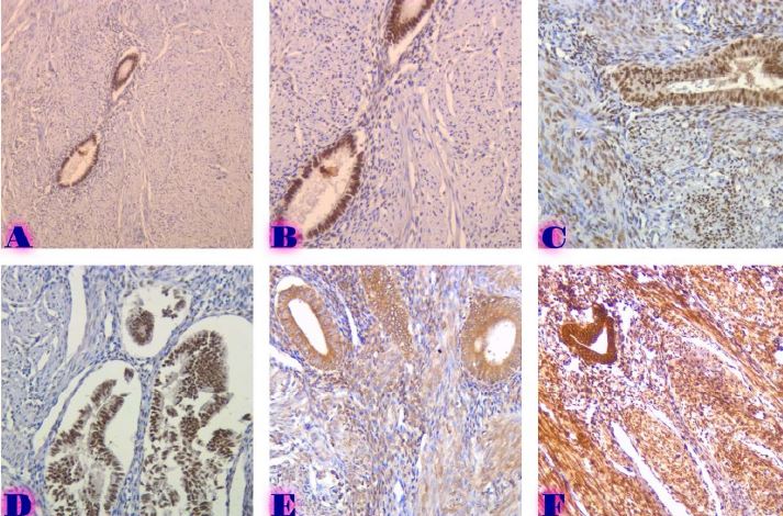
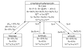
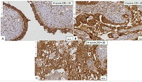
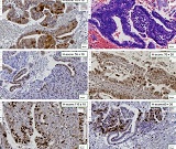
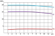
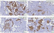
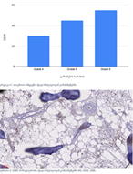
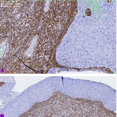
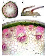
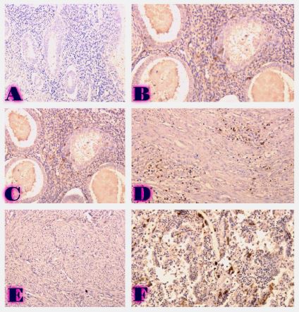
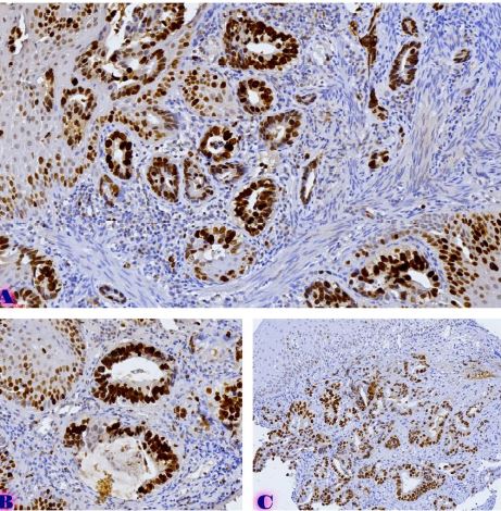
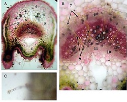
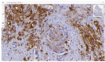
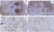
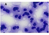
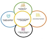
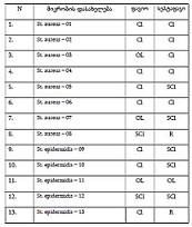
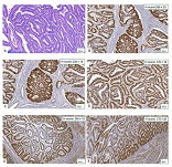
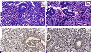
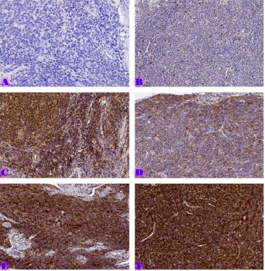
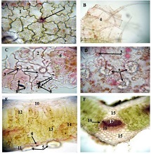
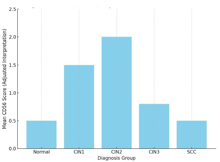
Despite the central role of epithelial dysplasia and HPV infection in cervical carcinogenesis, changes in the stromal microenvironment and neural remodelling remain the least studied areas. Our study examines the distribution and structural changes of peripheral nerve fibres across the spectrum of cervical intraepithelial neoplasia and invasive squamous cell carcinoma. Forty-five cervical tissue samples (9 normal, CIN1, CIN2, CIN3, and SCC) were stained using antibodies to PGP9.5, S100, CD56, and CD34. The density, maximum diameter, and distance from the epithelial basement membrane of nerve fibres were quantified. S100 and CD56 expression were semiquantitatively assessed. A progressive increase in PGP9.5-positive nerve fibres was observed from CIN1 to SCC, which was also accompanied by an increase in nerve fibre diameter. Hypertrophy of nerve fibres progressively increases with increasing dysplasia. Some fibres are normal, while others are neurogenic fibres resulting from neoangiogenesis. In CIN3 and SCC, NK (natural killer cells) are sharply reduced, which is explained by the presence of neoneurogenic fibres that dominate at this stage. Initially, in cases of low-grade dysplasia, NK cell hyperplasia increases along with hypertrophy, although its number is minimal in CIN3 and SCC. This may represent a manifestation of escape from neuroregulation/tumour escape from the immune system. These results may be used to refine immunotherapy better and develop NK-targeted drugs.
Downloads
მიმოხილვაკრიტიკული, გოგიტიძეგიორგი, კეპულაძეშოთა, თევზაძენინო, and ბურკაძეგიორგი, “The Role of the Local Neural microenvironment in the Progression of cervical intraepithelial Neoplasia,” Georgian Scientists, vol. 5, no. 3, pp. 182–188, Sep. 2023, doi: 10.52340/2023.05.03.19.
B. Boilly, S. Faulkner, P. Jobling, and H. Hondermarck, “Nerve Dependence: From Regeneration to Cancer,” Cancer Cell, vol. 31, no. 3, pp. 342–354, Mar. 2017, doi: 10.1016/J.CCELL.2017.02.005.
W. Wang et al., “Nerves in the Tumor Microenvironment: Origin and Effects,” Front Cell Dev Biol, vol. 8, Dec. 2020, doi: 10.3389/FCELL.2020.601738.
S. Faulkner, P. Jobling, B. March, C. C. Jiang, and H. Hondermarck, “Tumor neurobiology and the war of nerves in cancer,” Cancer Discov, vol. 9, no. 6, pp. 702–710, Jun. 2019, doi: 10.1158/2159-8290.CD-18-1398.
P. D. Vermeer, “Exosomal induction of tumor innervation,” Cancer Res, vol. 79, no. 14, pp. 3529–3535, 2019, doi: 10.1158/0008-5472.CAN-18-3995.
J. Bründl, S. Schneider, F. Weber, F. Zeman, W. F. Wieland, and R. Ganzer, “Computerized quantification and planimetry of prostatic capsular nerves in relation to adjacent prostate cancer foci,” Eur Urol, vol. 65, no. 4, pp. 802–808, Apr. 2014, doi: 10.1016/J.EURURO.2013.04.043.
C. T. Lucido et al., “Innervation of cervical carcinoma is mediated by cancer-derived exosomes,” Gynecol Oncol, vol. 154, no. 1, pp. 228–235, Jul. 2019, doi: 10.1016/J.YGYNO.2019.04.651.
L. Gao, H. Bo, Y. Wang, J. Zhang, and M. Zhu, “Neurotrophic factor Artemin promotes invasiveness and neurotrophic function of pancreatic Adenocarcinoma in vivo and in vitro,” Pancreas, vol. 44, no. 1, pp. 134–143, Jan. 2015, doi: 10.1097/MPA.0000000000000223.
S. Gillespie and M. Monje, “The Neural Regulation of Cancer,” Annu Rev Cancer Biol, vol. 4, pp. 371–390, Mar. 2020, doi: 10.1146/ANNUREV-CANCERBIO-030419-033349.
M. Arese, F. Bussolino, M. Pergolizzi, L. Bizzozero, and D. Pascal, “Tumor progression: the neuronal input,” Ann Transl Med, vol. 6, no. 5, pp. 89–89, Mar. 2018, doi: 10.21037/ATM.2018.01.01.
C. Magnon et al., “Autonomic nerve development contributes to prostate cancer progression,” Science (1979), vol. 341, no. 6142, 2013, doi: 10.1126/SCIENCE.1236361.
T. Dzotsenidze, A. Gvenetadze, M. Gachechiladze, G. Burkadze, and S. Kepuladze, “Immunohistochemical phenotype of fallopian tubes in patients with different grades of serous ovarian carcinoma,” Indian Journal of Pathology and Oncology Journal homepage: www.ijpo.co.in Original Research Article, vol. 9, no. 4, pp. 301–305, 2022, doi: 10.18231/j.ijpo.2022.073.
M. Głowienka-Stodolak et al., “Human Papillomavirus Infections and the Role Played by Cervical and Cervico-Vaginal Microbiota—Evidence from Next-Generation Sequencing Studies,” Cancers (Basel), vol. 16, no. 2, p. 399, Jan. 2024, doi: 10.3390/CANCERS16020399.
M. Schiffman and N. Wentzensen, “Human papillomavirus infection and the multistage carcinogenesis of cervical cancer,” Cancer Epidemiol Biomarkers Prev, vol. 22, no. 4, pp. 553–560, Apr. 2013, doi: 10.1158/1055-9965.EPI-12-1406.
Copyright (c) 2025 Georgian Scientists

This work is licensed under a Creative Commons Attribution-NonCommercial-NoDerivatives 4.0 International License.