დიაგნოსტიკური სირთულეები მაღალი ხარისხის ავთვისებიანობის პროსტატის ადენოკარცინომისა და მაღალი ხარისხის ავთვისებიანობის უროთელური კარცინომის თანაარსებობისას - ლიტერატურის მიმოხილვა
ჩამოტვირთვები
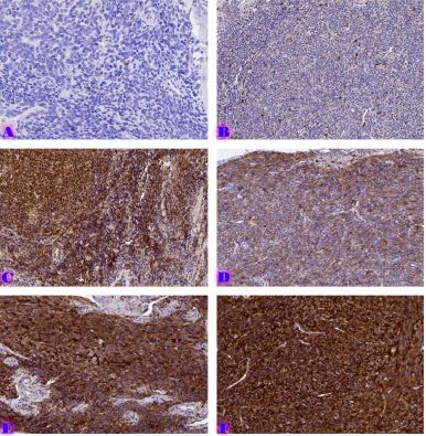
აბსტრაქტი: მამაკაცებში ორ ყველაზე გავრცელებულ სიმსივნეს მიეკუთვნება პროსტატის ადენოკარცინომა და გარდამავალუჯრედული, იგივე უროთელური კარცინომა. ორივე სიმსივნე შესაძლოა გამოვლინდეს ერთმანეთისაგან დამოუკიდებლად პროსტატაში ან შარდის ბუშტში ან ერთდროულად ცალკეულ სიმსივნეებად, რომლებიც ერთ-ერთ ორგანოშია გავრცელებული ან სპორადულად ე.წ. შერწყმული სიმსივნის სახით. პათოლოგანატომისათვის ამ სიმსივნეების ერთმანეთისაგან განსხვავება წარმოადგენს დიაგნოსტიკურ სირთულეს მაღალი ხარისხის ავთვისებიანობის მქონე ფორმების დროს, როდესაც ხდება სიმსივნის ირგვლივმდებარე ორგანოებში გავრცელება. აღნიშნული სიმსივნეების განსხვავებული მკურნალობის ტაქტიკის გამო მნიშვნელოვანია მათი სწორი დიაგნოსტიკა. ჩვენი მიმოხილვითი სტატიით შეკრებილია ინფორმაცია დიაგნოსტიკური სირთულეებისა და გადამწყვეტი იმუნოჰისტოქიმიური მარკერების შესახებ პროსტატის ადენოკარცინომისა და უროთელური კარცინომის სწორად დიფერენცირების მიზნით. ამ მიმოხილვაში გამოყენებულია PubMed და Google Scholar მონაცემთა ბაზების აკადემიურ ჟურნალებში გამოქვეყნებული სამეცნიერო ნაშრომები. მათგან მოპოვებულია პათოლოგანატომებისათვის მნიშვნელოვანი, აქტუალური და უახლესი ინფორმაცია. წარმოდგენილი მიმოხილვით გამოვლინდა რომ: 1) პროსტატის ადენოკარცინომისა და უროთელური კარცინომის დიფერენცირების მიზნით ყველაზე ინფორმატიული იმუნოჰისტოქიმიური მარკერებია: NKX3.1, p63, თრომბომოდულინი და GATA3. 2) პროსტატის სპეციფიკური ანტიგენი (PSA) არის კარგი კლინიკური სასკრინინგო მარკერი, თუმცა მაღალი ხარისხის ავთვისებიანობის მქონე სიმსივნეებში ის კარგავს სენსიტიურობას (რაც უფრო მაღალია ავთვისებიანობის ხარისხი, იგივე გრეიდი, მით უფრო დაბალია PSA მგრძნობელობა/შეღებვის ინტენსივობა) და სწორად ვერ ადიფერენცირებს აღწერილ სიმსივნეებს ერთმანეთისაგან. 3) უროთელური კარცინომის გამოსავლენად HMWCK (34βe12) და p63 უფრო ეფექტურია, ვიდრე თრომბომოდულინი და S100p. 4) თრომბომოდულინი გამოირჩევა ზომიერი მგრძნობელობით უროთელური კარცინომის დიაგნოსტიკაში. 5) ციტოკერატინი 7 და 20 შესაძლოა დადებითი იყოს როგორც უროთელური კარცინომის, ასევე პროსტატის ადენოკარცინომის დროს, ამიტომ მათი გამოყენება ნაკლებად სასარგებლო ინფორმაციის მომცემია. წარმოდგენილი მარკერების ოპტიმალური კომბინაციით შესაძლებელია გაუმჯობესებულ იქნას აღწერილი სიმსივნეების დიფერენციული დიაგნოსტიკა.
Downloads
Metrics
Mohanty SK, Smith SC, Chang E, et al. Evaluation of contemporary prostate and urothelial lineage biomarkers in a consecutive cohort of poorly differentiated bladder neck carcinomas. Am J Clin Pathol. 2014;142(2):173-183. doi:10.1309/AJCPK1OV6IMNPFGL
Oh WJ, Chung AM, Kim JS, et al. Differential Immunohistochemical Profiles for Distinguishing Prostate Carcinoma and Urothelial Carcinoma. J Pathol Transl Med. 2016;50(5):345-354. doi:10.4132/jptm.2016.06.14
Martínez-Rodríguez M, Ramos D, Soriano P, Subramaniam M, Navarro S, LlombartBosch A. Poorly differentiated adenocarcinomas of the prostate versus high- grade urothelial carcinoma of the bladder: a diagnostic dilemma with the immunohistochemical evaluation of 2 cases. Int J Surg Pathol. 2007;15(2):213-218. doi:10.1177/1066896906295822
Epstein JI, Egevad L, Humphrey PA, Montironi R; Members of the ISUP Immunohistochemistry in Diagnostic Urologic Pathology Group. Best practices recommendations in the application of immunohistochemistry in the prostate: report from the International Society of Urologic Pathology consensus conference. Am J Surg Pathol. 2014;38(8):e6-e19. doi:10.1097/PAS.0000000000000238
Signoretti S, Waltregny D, Dilks J, et al. p63 is a prostate basal cell marker and is required for prostate development. Am J Pathol. 2000;157(6):1769-1775. doi:10.1016/S00029440(10)64814-6
Amin MB, Trpkov K, Lopez-Beltran A, Grignon D; Members of the ISUP Immunohistochemistry in Diagnostic Urologic Pathology Group. Best practices recommendations in the application of immunohistochemistry in the bladder lesions: report from the International Society of Urologic Pathology consensus conference. Am J Surg Pathol. 2014;38(8):e20-e34. doi:10.1097/PAS.0000000000000240.
Genega EM, Hutchinson B, Reuter VE, Gaudin PB. Immunophenotype of high-grade prostatic adenocarcinoma and urothelial carcinoma. Mod Pathol. 2000;13(11):1186- 1191. doi:10.1038/modpathol.3880220
Chuang AY, DeMarzo AM, Veltri RW, Sharma RB, Bieberich CJ, Epstein JI. Immunohistochemical differentiation of high-grade prostate carcinoma from urothelial carcinoma. Am J Surg Pathol. 2007;31(8):1246-1255. doi:10.1097/PAS.0b013e31802f5d33.
Hameed O, Humphrey PA. Immunohistochemistry in diagnostic surgical pathology of the prostate. Semin Diagn Pathol. 2005;22(1):88-104. doi:10.1053/j.semdp.2005.11.001
Hameed O, Humphrey PA. Immunohistochemistry in diagnostic surgical pathology of the prostate. Semin Diagn Pathol. 2005;22(1):88-104. doi:10.1053/j.semdp.2005.11.001
Higgins JP, Kaygusuz G, Wang L, et al. Placental S100 (S100P) and GATA3: markers for transitional epithelium and urothelial carcinoma discovered by complementary DNA microarray. Am J Surg Pathol. 2007;31(5):673-680. doi:10.1097/01.pas.0000213438.01278.5f
Miettinen M, McCue PA, Sarlomo-Rikala M, et al. GATA3: a multispecific but potentially useful marker in surgical pathology: a systematic analysis of 2500 epithelial and nonepithelial tumors. Am J Surg Pathol. 2014;38(1):13-22. doi:10.1097/PAS.0b013e3182a0218f
Rubin MA, Zhou M, Dhanasekaran SM, et al. alpha-Methylacyl coenzyme A racemase as a tissue biomarker for prostate cancer. JAMA. 2002;287(13):1662-1670. doi:10.1001/jama.287.13.1662.
Luo J, Zha S, Gage WR, et al. Alpha-methyl acyl-CoA racemase: a new molecular marker for prostate cancer. Cancer Res. 2002;62(8):2220-2226.
Paner GP, Luthringer DJ, Amin MB. Best practice in diagnostic immunohistochemistry: prostate carcinoma and its mimics in needle core biopsies. Arch Pathol Lab Med. 2008;132(9):1388-1396. doi:10.5858/2008-132-1388-BPIDIP
Ud Din N, Qureshi A, Mansoor S. Utility of p63 immunohistochemical stain in differentiating urothelial carcinomas from adenocarcinomas of prostate. Indian J Pathol Microbiol. 2011;54(1):59-62. doi:10.4103/0377-4929.77326
Ordóñez NG. Thrombomodulin expression in transitional cell carcinoma. Am J Clin Pathol. 1998;110(3):385-390. doi:10.1093/ajcp/110.3.385
Mohammed KH, Siddiqui MT, Cohen C. GATA3 immunohistochemical expression in invasive urothelial carcinoma. Urol Oncol. 2016;34(10):432.e9-432.e13. doi:10.1016/j.urolonc.2016.04.016
McDonald TM, Epstein JI. Aberrant GATA3 Staining in Prostatic Adenocarcinoma: A Potential Diagnostic Pitfall. Am J Surg Pathol. 2021;45(3):341-346. doi:10.1097/PAS.0000000000001557.
Chou J, Provot S, Werb Z. GATA3 in development and cancer differentiation: cells GATA has it! J Cell Physiol. 2010;222(1):42-49. doi:10.1002/jcp.21943.
Miettinen M, McCue PA, Sarlomo-Rikala M, et al. GATA3: a multispecific but potentially useful marker in surgical pathology: a systematic analysis of 2500 epithelial and nonepithelial tumors. Am J Surg Pathol. 2014;38(1):13-22. doi:10.1097/PAS.0b013e3182a0218f
Tian W, Dorn D, Wei S, et al. GATA3 expression in benign prostate glands with radiation atypia: a diagnostic pitfall. Histopathology. 2017;71(1):150-155. doi:10.1111/his.13214
Olsburgh J, Harnden P, Weeks R, et al. Uroplakin gene expression in normal human tissues and locally advanced bladder cancer. J Pathol. 2003;199(1):41-49. doi:10.1002/path.1252
Kageyama S, Yoshiki T, Isono T, et al. High expression of human uroplakin Ia in urinary bladder transitional cell carcinoma. Jpn J Cancer Res. 2002;93(5):523-531. doi:10.1111/j.13497006.2002.tb01287.
Mhawech P, Uchida T, Pelte MF. Immunohistochemical profile of high-grade urothelial bladder carcinoma and prostate adenocarcinoma. Hum Pathol. 2002;33(11):1136-1140. doi:10.1053/hupa.2002.129416
Xu X, Sun TT, Gupta PK, Zhang P, Nasuti JF. Uroplakin as a marker for typing metastatic transitional cell carcinoma on fine-needle aspiration specimens. Cancer. 2001;93(3):216- 221. doi:10.1002/cncr.9032
Chuang AY, DeMarzo AM, Veltri RW, Sharma RB, Bieberich CJ, Epstein JI. Immunohistochemical differentiation of high-grade prostate carcinoma from urothelial carcinoma. Am J Surg Pathol. 2007;31(8):1246-1255. doi:10.1097/PAS.0b013e31802f5d33

ეს ნამუშევარი ლიცენზირებულია Creative Commons Attribution-NonCommercial-NoDerivatives 4.0 საერთაშორისო ლიცენზიით .


