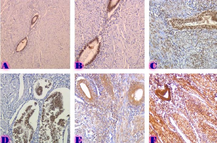
Histological and immunohistochemical profile of lymphocytic duodenitis and features of local immune reactions
Downloads
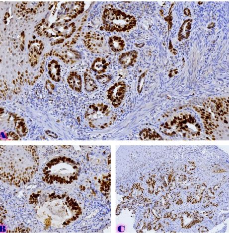
Lymphocytic enteritis is characterised by abnormal infiltration of intraepithelial lymphocytes in the intestinal mucosa. It was described as duodenal lymphocytosis or lymphocytic duodenitis. Nowadays, lymphocytic enteritis represents a common feature of several gluten-mediated and non-gluten-related diseases. The most frequent causes of lymphocytic enteritis are gluten-related disorders (celiac disease, non-celiac gluten sensitivity), Helicobacter pylori infection and drug-related damages. Less frequently, lymphocytic enteritis may be secondary to autoimmune conditions, immunoglobulin deficiencies, bacterial and viral infections and irritable bowel syndrome. Therefore, the differential diagnosis of lymphocytic enteritis may be challenging.
Downloads
G. Caio et al., “Celiac disease: a comprehensive current review.,” BMC Med, vol. 17, no. 1, p. 142, Jul. 2019, doi: 10.1186/s12916-019-1380-z.
S. Husby, J. A. Murray, and D. A. Katzka, “AGA Clinical Practice Update on Diagnosis and Monitoring of Celiac Disease-Changing Utility of Serology and Histologic Measures: Expert Review.,” Gastroenterology, vol. 156, no. 4, pp. 885–889, Mar. 2019, doi: 10.1053/j.gastro.2018.12.010.
J. A. Silvester, A. Therrien, and C. P. Kelly, “Celiac Disease: Fallacies and Facts.,” Am J Gastroenterol, vol. 116, no. 6, pp. 1148–1155, Jun. 2021, doi: 10.14309/ajg.0000000000001218.
V. Villanacci et al., “Celiac disease: histology-differential diagnosis-complications. A practical approach.,” Pathologica, vol. 112, no. 3, pp. 186–196, Sep. 2020, doi: 10.32074/1591-951X-157.
V. Villanacci et al., “Celiac disease: histology-differential diagnosis-complications. A practical approach.,” Pathologica, vol. 112, no. 3, pp. 186–196, Sep. 2020, doi: 10.32074/1591-951X-157.
R. Del Sordo et al., “Histological Features of Celiac-Disease-like Conditions Related to Immune Checkpoint Inhibitors Therapy: A Signal to Keep in Mind for Pathologists.,” Diagnostics (Basel), vol. 12, no. 2, Feb. 2022, doi: 10.3390/diagnostics12020395.
R. Dieli-Crimi, M. C. Cénit, and C. Núñez, “The genetics of celiac disease: A comprehensive review of clinical implications.,” J Autoimmun, vol. 64, pp. 26–41, Nov. 2015, doi: 10.1016/j.jaut.2015.07.003.
A. K. Akobeng, P. Singh, M. Kumar, and S. Al Khodor, “Role of the gut microbiota in the pathogenesis of coeliac disease and potential therapeutic implications.,” Eur J Nutr, vol. 59, no. 8, pp. 3369–3390, Dec. 2020, doi: 10.1007/s00394-020-02324-y.
J. A. Tye-Din, H. J. Galipeau, and D. Agardh, “Celiac Disease: A Review of Current Concepts in Pathogenesis, Prevention, and Novel Therapies.,” Front Pediatr, vol. 6, p. 350, 2018, doi: 10.3389/fped.2018.00350.
G. Caio et al., “Celiac disease: a comprehensive current review.,” BMC Med, vol. 17, no. 1, p. 142, Jul. 2019, doi: 10.1186/s12916-019-1380-z.
E. M. Quinn et al., “Transcriptome Analysis of CD4+ T Cells in Coeliac Disease Reveals Imprint of BACH2 and IFNγ Regulation.,” PLoS One, vol. 10, no. 10, p. e0140049, 2015, doi: 10.1371/journal.pone.0140049.
R. Celli et al., “Clinical Insignficance of Monoclonal T-Cell Populations and Duodenal Intraepithelial T-Cell Phenotypes in Celiac and Nonceliac Patients.,” Am J Surg Pathol, vol. 43, no. 2, pp. 151–160, Feb. 2019, doi: 10.1097/PAS.0000000000001172.
Y. Sahin, “Celiac disease in children: A review of the literature.,” World J Clin Pediatr, vol. 10, no. 4, pp. 53–71, Jul. 2021, doi: 10.5409/wjcp.v10.i4.53.
P. Laurikka, L. Kivelä, K. Kurppa, and K. Kaukinen, “Review article: Systemic consequences of coeliac disease.,” Aliment Pharmacol Ther, vol. 56 Suppl 1, no. Suppl 1, pp. S64–S72, Jul. 2022, doi: 10.1111/apt.16912.
S. Hussein et al., “Clonal T cell receptor gene rearrangements in coeliac disease: implications for diagnosing refractory coeliac disease.,” J Clin Pathol, vol. 71, no. 9, pp. 825–831, Sep. 2018, doi: 10.1136/jclinpath-2018-205023.
A. Lerner, M. Arleevskaya, A. Schmiedl, and T. Matthias, “Microbes and Viruses Are Bugging the Gut in Celiac Disease. Are They Friends or Foes?,” Front Microbiol, vol. 8, p. 1392, 2017, doi: 10.3389/fmicb.2017.01392.
G. Caio, G. Riegler, M. Patturelli, A. Facchiano, L. DE Magistris, and A. Sapone, “Pathophysiology of non-celiac gluten sensitivity: where are we now?,” Minerva Gastroenterol Dietol, vol. 63, no. 1, pp. 16–21, Mar. 2017, doi: 10.23736/S1121-421X.16.02346-1.
V. F. Zevallos et al., “Nutritional Wheat Amylase-Trypsin Inhibitors Promote Intestinal Inflammation via Activation of Myeloid Cells.,” Gastroenterology, vol. 152, no. 5, pp. 1100-1113.e12, Apr. 2017, doi: 10.1053/j.gastro.2016.12.006.
G. Losurdo et al., “Small intestinal bacterial overgrowth and celiac disease: A systematic review with pooled-data analysis.,” Neurogastroenterology and motility, vol. 29, no. 6, Jun. 2017, doi: 10.1111/nmo.13028.
M. Rostami-Nejad et al., “Pathological and Clinical Correlation between Celiac Disease and Helicobacter Pylori Infection; a Review of Controversial Reports.,” Middle East J Dig Dis, vol. 8, no. 2, pp. 85–92, Apr. 2016, doi: 10.15171/mejdd.2016.12.
A. K. Kamboj and A. S. Oxentenko, “Clinical and Histologic Mimickers of Celiac Disease.,” Clin Transl Gastroenterol, vol. 8, no. 8, p. e114, Aug. 2017, doi: 10.1038/ctg.2017.41.
D. C. Baumgart and C. Le Berre, “Newer Biologic and Small-Molecule Therapies for Inflammatory Bowel Disease.,” N Engl J Med, vol. 385, no. 14, pp. 1302–1315, Sep. 2021, doi: 10.1056/NEJMra1907607.
E. Ierardi et al., “Lymphocytic duodenitis or microscopic enteritis and gluten-related conditions: what needs to be explored?,” Ann Gastroenterol, vol. 30, no. 4, pp. 380–392, 2017, doi: 10.20524/aog.2017.0165.
Copyright (c) 2023 GEORGIAN SCIENTISTS

This work is licensed under a Creative Commons Attribution-NonCommercial-NoDerivatives 4.0 International License.


