
Histomorphological Heterogeneity of Melanocytic Nevi: Clinicopathological Features and Assessment of Progression Potential

Downloads




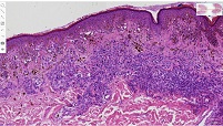
Melanocytic nevi are the most common benign melanocytic lesions of the skin, which are characterised by significant histomorphological diversity. The assessment of architectural, cytological and stromal features of different histological types of nevi is critically important, both in differential diagnosis and in better understanding their biological behaviour.
The study aimed to assess the histomorphological heterogeneity of different histological types of melanocytic nevi, compare their clinicopathological features and identify morphological signs associated with progression. The retrospective study included histological material from 52 patients diagnosed with different types of melanocytic nevi. Hematoxylin-eosin (H&E) stained slides were used to assess the histological type of nevus, symmetry, circumscription, maturation, bordering component, Pagetoid spread, cytological atypia, inflammatory infiltrate, fibrosis, solar elastosis, pigmentation, ulcerative changes, and resection margin status. Data were analysed using IBM SPSS Statistics 29.0.
The study showed that melanocytic nevi of different histological types differed significantly in architectural and stromal characteristics. Bordering, intradermal, compound, and blue nevi were mostly symmetrical and well-circumscribed. In contrast, dysplastic and Spitz nevi were more often characterised by asymmetrical architecture, partial maturation, cytological atypia, and inflammatory changes. Recurrences were mainly associated with the dysplastic nevus group and were more common in cases with positive surgical resection margins.
Melanocytic nevi are characterised by marked histomorphological heterogeneity, which depends on their histological type. Routine histomorphological evaluation provides important information on clinicopathological features and morphological signs of progression and serves as a basis for subsequent immunohistochemical and digital pathology studies.
Downloads
[1] Daltro LR, Freitas RA, Souza C da S, Yaegashi LB, Fantini B de C. Atypical cellular blue nevus or malignant blue nevus? An Bras Dermatol 2017;92:110. https://doi.org/10.1590/ABD1806-4841.20174502.
[2] Zalaudek I, Conforti C, Guarneri F, Vezzoni R, Deinlein T, Hofmann-Wellenhof R, et al. Clinical and dermoscopic characteristics of congenital and noncongenital nevus-associated melanomas. J Am Acad Dermatol 2020;83:1080–7. https://doi.org/10.1016/J.JAAD.2020.04.120.
[3] Yeh I, Busam KJ. Spitz melanocytic tumours – a review. Histopathology 2022;80:122–34. https://doi.org/10.1111/HIS.14583.
[4] Mccarthy JM, Rootman J, Horsman D, White VA. Conjunctival and uveal melanoma in the dysplastic nevus syndrome. Surv Ophthalmol 1993;37:377–86. https://doi.org/10.1016/0039-6257(93)90068-I.
[5] Seregard S, Trampe E af, Månsson-Brahme E, Kock E, Bergenmar M, Ringborg U. Prevalence of Primary Acquired Melanosis and Nevi of the Conjunctiva and Uvea in the Dysplastic Nevus Syndrome: A Case-control Study. Ophthalmology 1995;102:1524–9. https://doi.org/10.1016/S0161-6420(95)30836-6 .
[6] Li WJ, Venkateswaran N, Antonietti M, Qureshi S, Galor A, Karp CL. Emerging Techniques in the Treatment of Conjunctival Melanoma. Curr Ophthalmol Rep 2025;13:7. https://doi.org/10.1007/s40135-025-00334-9.
[7] Shain AH, Bastian BC. From melanocytes to melanomas. Nat Rev Cancer 2016;16:345–58. https://doi.org/10.1038/NRC.2016.37.
[8] Salvati L, Palmieri G, De Giorgi V, Cossu A, Mandalà M, Massi D. Melanoma: Pathology and genetics. Encyclopedia of Cancer 2018:414–25. https://doi.org/10.1016/B978-0-12-801238-3.65095-X.
[9] de la Fouchardière A, Boivin F, Etchevers HC, Macagno N. Cutaneous Melanomas Arising during Childhood: An Overview of the Main Entities. Dermatopathology 2021;8:301–14. https://doi.org/10.3390/DERMATOPATHOLOGY8030036.
[10] Zalaudek I, Conforti C, Guarneri F, Vezzoni R, Deinlein T, Hofmann-Wellenhof R, et al. Clinical and dermoscopic characteristics of congenital and noncongenital nevus-associated melanomas. J Am Acad Dermatol 2020;83:1080–7. https://doi.org/10.1016/J.JAAD.2020.04.120.
[11] Pampena R, Lai M, Piana S, Lallas A, Pellacani G, Longo C. Nevus-associated melanoma: Facts and controversies. Giornale Italiano Di Dermatologia e Venereologia 2020;155:65–75. https://doi.org/10.23736/S0392-0488.19.06534-9.
[12] Dessinioti C, Geller AC, Stratigos AJ. A review of nevus-associated melanoma: What is the evidence? Journal of the European Academy of Dermatology and Venereology 2022;36:1927–36. https://doi.org/10.1111/jdv.18453.
Copyright (c) 2026 Georgian Scientists

This work is licensed under a Creative Commons Attribution-NonCommercial-NoDerivatives 4.0 International License.