Comparative analysis of phenotypic features in the primary tumor, tumour buds and metastatic lymph nodes within Luminal A and Luminal B molecular subtypes of invasive ductal carcinoma of the breast
Downloads
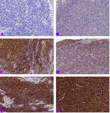
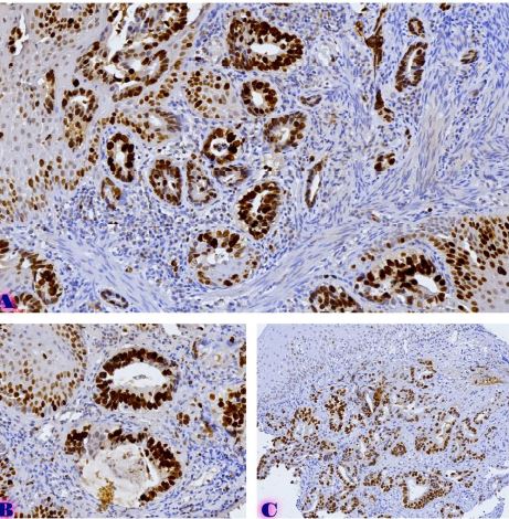
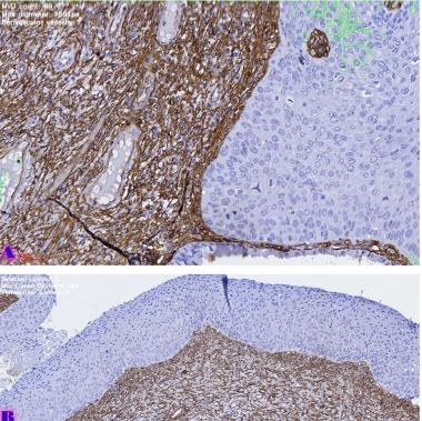
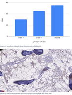
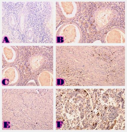
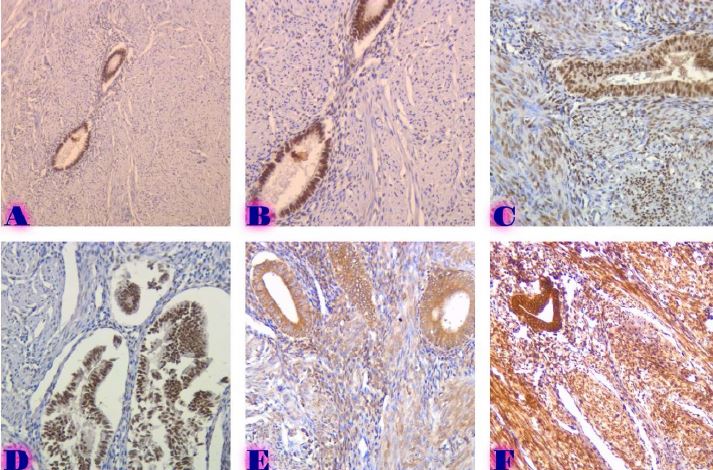
The study included 55 cases of formalin-fixed and paraffin-embedded (FFPE) tissues of breast invasive ductal carcinoma. The following algorithm has been made for further discussion by using immunohistochemical examination: antibodies against hormonal receptors; HER2; E-cadherin, Vimentin, Beta-catenin; Ki-67; Tumour buds were evaluated by using H&E stained slides and computer software Qupath (version 0.3.2). The results of the study show that estrogen expression is different in primary tumour mass and in tumour buds and its expression is diminished in the Luminal B molecular subtype respectively. Vimentin and Beta-catenin expression is showing similar changes, the quantity is much higher in tumour microclusters compared to the primary tumour and metastatic lymph nodes. It can demonstrate features of epithelial-mesenchymal transformation. Modifications in dynamics of proliferative activity are showing the lowest proliferative activity in tumour microclusters which can be discussed as the indirect manifestation of epithelial-mesenchymal transformation.
Downloads
R. L. Siegel, K. D. Miller, and A. Jemal, “Cancer statistics, 2019,” CA: A Cancer Journal for Clinicians, vol. 69, no. 1, pp. 7–34, Jan. 2019, doi: 10.3322/CAAC.21551.
O. Okcu, Ç. Öztürk, B. Şen, M. Arpa, and R. Bedir, “Tumor Budding is a reliable predictor for death and metastasis in invasive ductal breast cancer and correlates with other prognostic clinicopathological parameters,” Annals of Diagnostic Pathology, vol. 54, Oct. 2021, doi: 10.1016/j.anndiagpath.2021.151792.
F. J. A. Gujam, D. C. McMillan, Z. M. A. Mohammed, J. Edwards, and J. J. Going, “The relationship between tumour budding, the tumour microenvironment and survival in patients with invasive ductal breast cancer,” British Journal of Cancer, vol. 113, no. 7, pp. 1066–1074, Sep. 2015, doi: 10.1038/BJC.2015.287.
B. Salhia et al., “High tumor budding stratifies breast cancer with metastatic properties,” Breast Cancer Research and Treatment, vol. 150, no. 2, pp. 363–371, Apr. 2015, doi: 10.1007/s10549-015-3333-3.
R. Agarwal, N. Khurana, T. Singh, and P. Agarwal, “Tumor budding in infiltrating breast carcinoma: Correlation with known clinicopathological parameters and hormone receptor status,” Indian Journal of Pathology and Microbiology, vol. 62, no. 2, p. 222, Apr. 2019, doi: 10.4103/IJPM.IJPM_120_18.
F. Liang, W. Cao, Y. Wang, L. Li, G. Zhang, and Z. Wang, “The prognostic value of tumor budding in invasive breast cancer,” Pathology Research and Practice, vol. 209, no. 5, pp. 269–275, May 2013, doi: 10.1016/j.prp.2013.01.009.
F. Liang, W. Cao, Y. Wang, L. Li, G. Zhang, and Z. Wang, “The prognostic value of tumor budding in invasive breast cancer,” Pathology Research and Practice, vol. 209, no. 5, pp. 269–275, May 2013, doi: 10.1016/j.prp.2013.01.009.
B. Salhia et al., “High tumor budding stratifies breast cancer with metastatic properties,” Breast Cancer Research and Treatment, vol. 150, no. 2, pp. 363–371, Apr. 2015, doi: 10.1007/S10549-015-3333-3.
B. N. Kumarguru, A. S. Ramaswamy, S. Shaik, A. Karri, V. S. Srinivas, and B. M. Prashant, “Tumor budding in invasive breast cancer - An indispensable budding touchstone,” Indian J Pathol Microbiol, vol. 63, no. Supplement, pp. S117–S122, Feb. 2020, doi: 10.4103/IJPM.IJPM_731_18.
J. van Staalduinen, D. Baker, P. ten Dijke, and H. van Dam, “Epithelial–mesenchymal-transition-inducing transcription factors: new targets for tackling chemoresistance in cancer?,” Oncogene, vol. 37, no. 48, pp. 6195–6211, Nov. 2018, doi: 10.1038/S41388-018-0378-X.
Y. Wu, M. Sarkissyan, and J. v. Vadgama, “Epithelial-mesenchymal transition and breast cancer,” Journal of Clinical Medicine, vol. 5, no. 2, pp. 1–18, Jan. 2016, doi: 10.3390/JCM5020013.
A. Dongre et al., “Epithelial-to-mesenchymal transition contributes to immunosuppression in breast carcinomas,” Cancer Research, vol. 77, no. 15, pp. 3982–3989, Aug. 2017, doi: 10.1158/0008-5472.CAN-16-3292.
M. Matadamas-Guzman, C. Zazueta, E. Rojas, and O. Resendis-Antonio, “Analysis of Epithelial-Mesenchymal Transition Metabolism Identifies Possible Cancer Biomarkers Useful in Diverse Genetic Backgrounds,” Frontiers in Oncology, vol. 10, Jul. 2020, doi: 10.3389/FONC.2020.01309/FULL.
S. Y. Cheung et al., “Role of epithelial–mesenchymal transition markers in triple-negative breast cancer,” Breast Cancer Research and Treatment, vol. 152, no. 3, pp. 489–498, Aug. 2015, doi: 10.1007/S10549-015-3485-1.
V. Mittal, “Epithelial Mesenchymal Transition in Tumor Metastasis,” Annual Review of Pathology: Mechanisms of Disease, vol. 13, pp. 395–412, Jan. 2018, doi: 10.1146/ANNUREV-PATHOL-020117-043854.
I. Zlobec and A. Lugli, “Epithelial mesenchymal transition and tumor budding in aggressive colorectal cancer: tumor budding as oncotarget.,” Oncotarget, vol. 1, no. 7, pp. 651–661, 2010, doi: 10.18632/ONCOTARGET.199.
I. Pastushenko and C. Blanpain, “EMT Transition States during Tumor Progression and Metastasis,” Trends in Cell Biology, vol. 29, no. 3, pp. 212–226, Mar. 2019, doi: 10.1016/J.TCB.2018.12.001.
N. Harbeck, C. Thomssen, and M. Gnant, “St. Gallen 2013: Brief Preliminary Summary of the Consensus Discussion,” 2013, doi: 10.1159/000351193.
P. Bankhead et al., “QuPath: Open source software for digital pathology image analysis,” Scientific Reports, vol. 7, no. 1, Dec. 2017, doi: 10.1038/S41598-017-17204-5.
A. Lugli et al., “Recommendations for reporting tumor budding in colorectal cancer based on the International Tumor Budding Consensus Conference (ITBCC) 2016,” Modern Pathology 2017 30:9, vol. 30, no. 9, pp. 1299–1311, May 2017, doi: 10.1038/modpathol.2017.46.
K. A. Bhuvaneshwari, “Expression of vimentin in breast carcinoma and its correlation with histopathological parameters,” Indian Journal of Pathology and Oncology Journal homepage: www.ijpo.co.in Original Research Article, vol. 8, no. 2, pp. 207–212, 2021, doi: 10.18231/j.ijpo.2021.041.

This work is licensed under a Creative Commons Attribution-NonCommercial-NoDerivatives 4.0 International License.


