
Four Modifiable Levels of Replicative Capacity: Centrioles, Progenitors, Gametes, Nutrition

Downloads




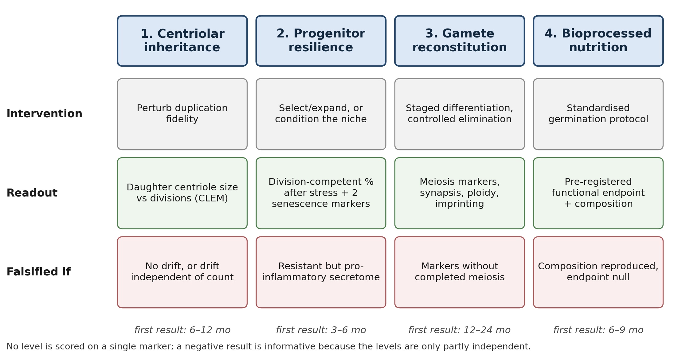 Ageing research is rich in candidate mechanisms and poor in operational readouts. The hallmarks framework organises damage into a coherent list [1,2], yet a laboratory that wants to slow replicative decline still has to choose which quantity it will actually measure, and a list of twelve mechanisms does not indicate which one should be perturbed first. We argue that replicative capacity - the number of divisions a cell lineage can complete before it loses function - is set at four partly independent levels, and that each level has a distinct, already available intervention and a distinct way of being wrong. The four are: structural inheritance of the mother centriole, resilience of tissue-resident progenitors to senescence, reconstitution of gametes from somatic cells, and bioprocess-based modification of nutrition. For each level we state three things: the intervention, the readout that would count as success, and the observation that would falsify the claim. That triad is what separates a declaration from an experiment. Our operational definition of replicative capacity is deliberately narrower than “healthspan” and wider than the Hayflick limit: it is measured by population doubling level, clone size distribution, or the fraction of cells retaining a lineage marker after serial passage, and it excludes organism-level endpoints, which we do not address. We then give a single multicentre-style test matrix that could be run with existing methods, with a realistic time to a first result for each level, and we add two design rules: no level is scored on a marker alone, and a negative result at one level is informative rather than fatal because the levels are claimed to be partly independent - which is also the matrix’s main vulnerability, since partial independence is easy to assert and hard to demonstrate. We are also explicit about how thin the published evidence base still is at three of the four levels, and about the self-citation that enters through the authors’ own institution. The purpose of the paper is not to propose a new theory of ageing; it is to make the existing claims testable in the same units.
Downloads
1. López-Otín, C.; Blasco, M.A.; Partridge, L.; Serrano, M.; Kroemer, G. Hallmarks of aging: An expanding universe. Cell 2023, 186, 243–278. doi:10.1016/j.cell.2022.11.001
2. López-Otín, C.; Blasco, M.A.; Partridge, L.; Serrano, M.; Kroemer, G. The hallmarks of aging. Cell 2013, 153, 1194–1217. doi:10.1016/j.cell.2013.05.039
3. Gorgoulis, V.; Adams, P.D.; Alimonti, A.; Bennett, D.C.; Bischof, O.; Bishop, C.; et al. Cellular senescence: Defining a path forward. Cell 2019, 179, 813–827. doi:10.1016/j.cell.2019.10.005
4. Zhu, Y.; Tchkonia, T.; Fuhrmann-Stroissnigg, H.; Dai, H.M.; Ling, Y.Y.; Stout, M.B.; et al. Identification of a novel senolytic agent, navitoclax, targeting the Bcl-2 family of anti-apoptotic factors. Aging Cell 2016, 15, 428–435. doi:10.1111/acel.12445
5. Kirkland, J.L.; Tchkonia, T. Senolytic drugs: From discovery to translation. J. Intern. Med. 2020, 288, 518–536. doi:10.1111/joim.13141
6. Horvath, S. DNA methylation age of human tissues and cell types. Genome Biol. 2013, 14, R115. doi:10.1186/gb-2013-14-10-r115
7. Nigg, E.A.; Holland, A.J. Once and only once: Mechanisms of centriole duplication and their deregulation in disease. Nat. Rev. Mol. Cell Biol. 2018, 19, 297–312. doi:10.1038/nrm.2017.127
8. Nigg, E.A.; Stearns, T. The centrosome cycle: Centriole biogenesis, duplication and inherent asymmetries. Nat. Cell Biol. 2011, 13, 1154–1160. doi:10.1038/ncb2345
9. Fu, J.; Hagan, I.M.; Glover, D.M. The centrosome and its duplication cycle. Cold Spring Harb. Perspect. Biol. 2015, 7, a015800. doi:10.1101/cshperspect.a015800
10. Katajisto, P.; Döhla, J.; Chaffer, C.L.; Pentinmikko, N.; Marjanovic, N.; Iqbal, S.; et al. Asymmetric apportioning of aged mitochondria between daughter cells is required for stemness. Science 2015, 348, 340–343. doi:10.1126/science.1260384
11. Gegelia, L. Mesenchymal progenitor cell resilience counteracts tissue aging. Longevity Horizon 2026, 2(1). doi:10.65649/c833dn40
12. Gegelia, L. A senescence-resistant mesenchymal progenitor cell sustains tissue homeostasis in ageing. Annals of Rejuvenation Science 2026, 1(1). doi:10.65649/cnxbvp22
13. Taktakidze, A. Engineering oocytes from somatic cells. Longevity Horizon 2026, 2(1). doi:10.65649/2q035e46
14. Gakely, G. Centriole regulation in natural and artificial gametogenesis. Longevity Horizon 2026, 2(1). doi:10.65649/87tnc168
15. Gakely, G. Centriole elimination in natural vs. in vitro gametogenesis. Annals of Rejuvenation Science 2026, 1(1). doi:10.65649/rnca4y67
16. Tqemaladze, J. Daughter centrioles are not chemical copies of their mothers: Geometric persistence as a replicative-age counter. Cytoskeleton 2026, e70222. doi:10.1002/cm.70222
17. Tkemaladze, J.; Gakely, G. A novel biotechnological approach for the production of shelf-stable, nutritionally enhanced activated wheat: Protocol development, nutritional profiling, and bioactivity assessment. Preprints 2025, 2025081997. doi:10.20944/preprints202508.1997.v1
18. Saitou, M.; Hayashi, K. Mammalian germ cell development: From mechanism to in vitro reconstitution. Stem Cell Rep. 2021, 16, 669–680. doi:10.1016/j.stemcr.2021.01.008
19. Hayashi, K. In vitro reconstitution of germ cell development. Biol. Reprod. 2019, 101, 567–578. doi:10.1093/biolre/ioz111
20. Rando, T.A.; Brunet, A.; Goodell, M.A. Hallmarks of stem cell aging. Cell Stem Cell 2025, 32, 1038–1054. doi:10.1016/j.stem.2025.06.004
21. Hickson, L.J.; Langhi Prata, L.G.P.; Bobart, S.A.; Evans, T.K.; Giorgadze, N.; Hashmi, S.K.; et al. Senolytics decrease senescent cells in humans: Preliminary report from a clinical trial of dasatinib plus quercetin in individuals with diabetic kidney disease. EBioMedicine 2019, 47, 446–456. doi:10.1016/j.ebiom.2019.08.069
22. Jiang, X.; Li, W.; Ge, L.; Lu, M. Mesenchymal stem cell senescence during aging: From mechanisms to rejuvenation strategies. Aging Dis. 2023, 14, 1651–1676. doi:10.14336/ad.2023.0208
23. Liu, J.; Ding, Y.; Liu, Z.; Liang, X. Senescence in mesenchymal stem cells: Functional alterations, molecular mechanisms, and rejuvenation strategies. Front. Cell Dev. Biol. 2020, 8, 258. doi:10.3389/fcell.2020.00258
24. Kalbfuss, N.; Gönczy, P. Towards understanding centriole elimination. Open Biol. 2023, 13, 230222. doi:10.1098/rsob.230222
25. Pimenta-Marques, A.; Bento, I.; Lopes, C.A.M.; Duarte, P.; Jana, S.C.; Bettencourt-Dias, M. A mechanism for the elimination of the female gamete centrosome in Drosophila melanogaster. Science 2016, 353, aaf4866. doi:10.1126/science.aaf4866
26. Hung, P.V.; Trinh, L.N.D.; Thuy, N.T.X. Changes in nutritional composition, enzyme activities and bioactive compounds of germinated wheat. Int. J. Food Sci. Technol. 2021, 56(7), 3209–3217 (online 22 November 2020). doi:10.1111/ijfs.14883
Note on transliteration. References 16 and 17 name the same co-author, Jaba Tqemaladze (ORCID 0000-0001-8651-7243). The two spellings are exactly those registered in the metadata of the respective publishers — Tqemaladze for the Cytoskeleton article (10.1002/cm.70222) and Tkemaladze for the Preprints.org record (10.20944/preprints202508.1997.v1) — and each entry has been kept as published so that both resolve to the deposited record.
Copyright (c) 2026 Georgian Scientists

This work is licensed under a Creative Commons Attribution-NonCommercial-NoDerivatives 4.0 International License.