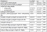
მელატონინის რეცეპტორების ექსპრესიის თავისებურებები სხვადასხვა ტიპის კოლორექტულ პოლიპებში, მისი კავშირი პროლიფერაციულ-აპოპტოზური, ჰორმონული რეცეპტორების ექსპრესიასა და ღეროვან უჯრედულ მახასიათებლებთან

ჩამოტვირთვები


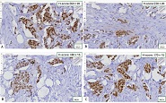
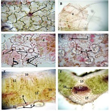
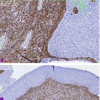
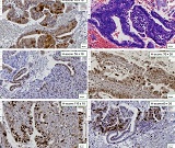
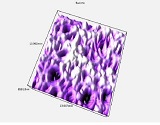
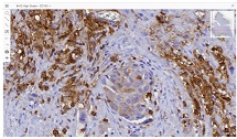
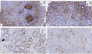
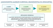
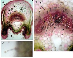
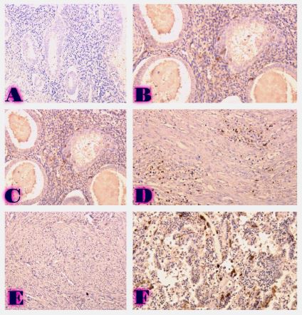
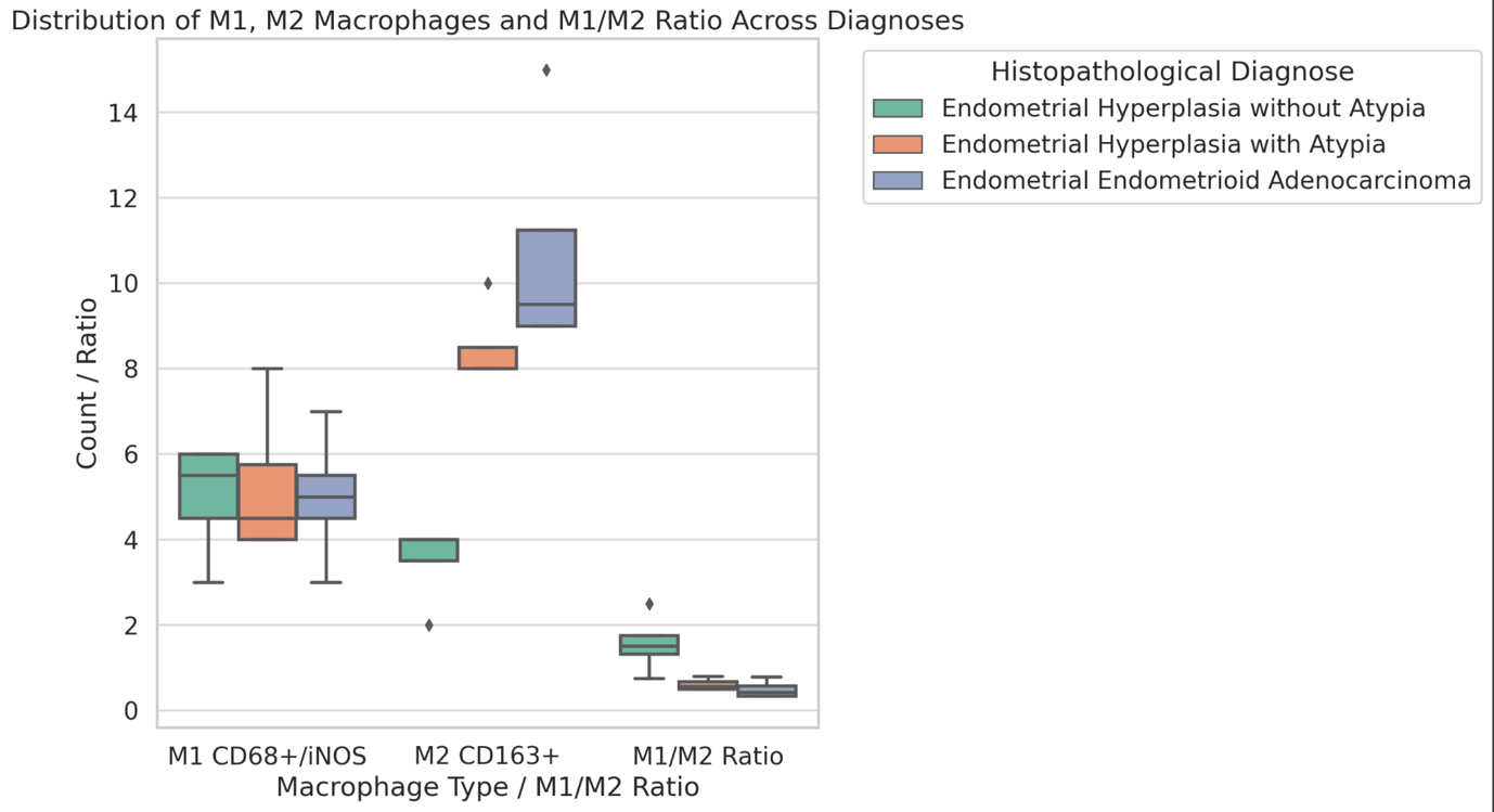
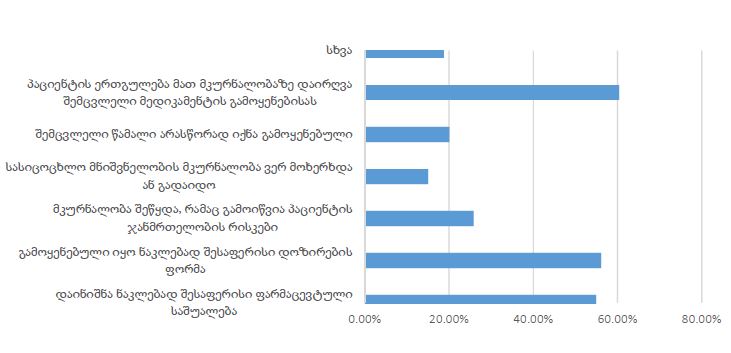
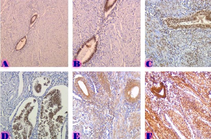
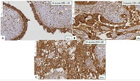
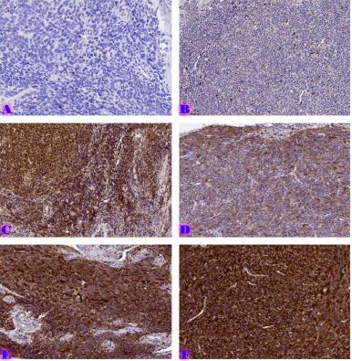
კოლორექტული პოლიპები წარმოადგენს ჰეტეროგენულ ჯგუფს, რომელიც მოიცავს როგორც კეთილთვისებიან, ასევე კიბოსწინარე დაზიანებებს და მნიშვნელოვან როლს ასრულებს კოლორექტული კარცინოგენეზის პროცესში. ბოლო წლებში განსაკუთრებული ყურადღება ეთმობა მელატონინისა და მისი რეცეპტორების (MT1, MT2) მონაწილეობას სიმსივნური პროცესების რეგულაციაში. მელატონინის რეცეპტორები დაკავშირებულია უჯრედული პროლიფერაციის, აპოპტოზის, ანგიოგენეზის, იმუნური პასუხისა და ღეროვანი უჯრედების ბიოლოგიის კონტროლთან, რაც მათ პერსპექტიულ ბიომარკერებად აქცევს სხვადასხვა ნეოპლაზიური პროცესის შეფასებისათვის.
წინამდებარე ლიტერატურული მიმოხილვის მიზანია კოლორექტულ პოლიპებში მელატონინის რეცეპტორების ექსპრესიის თანამედროვე მონაცემების ანალიზი და მათი კავშირის შეფასება დისპლაზიის ხარისხთან, პროლიფერაციულ-აპოპტოზურ აქტივობასთან, ჰორმონული რეცეპტორების ექსპრესიასა და ღეროვანი უჯრედების მახასიათებლებთან. განხილულია MT1 და MT2 რეცეპტორების ბიოლოგიური როლი, მათი მონაწილეობა Wnt/β-კატენინის, MAPK/ERK და სხვა სასიგნალო გზების რეგულაციაში, აგრეთვე მათი გავლენა კოლორექტული ნეოპლაზიის განვითარებასა და პროგრესირებაზე.
ლიტერატურაში არსებული მონაცემები მიუთითებს, რომ მელატონინის რეცეპტორების ექსპრესიის შემცირება შეიძლება ასოცირებული იყოს დისპლაზიის პროგრესირებასთან, უჯრედული პროლიფერაციის გაძლიერებასთან, აპოპტოზის დათრგუნვასთან და უფრო აგრესიული ბიოლოგიური ქცევის ფორმირებასთან. ასევე სავარაუდოა მათი კავშირი ჰორმონული რეგულაციისა და ღეროვანი უჯრედების მარკერებთან, რაც დამატებით უსვამს ხაზს მელატონინის სასიგნალო სისტემის მნიშვნელობას კოლორექტალული კარცინოგენეზის პროცესში.
მელატონინის რეცეპტორების დეტალური შესწავლა შეიძლება გახდეს ახალი პროგნოზული ბიომარკერების იდენტიფიკაციის საფუძველი და ხელი შეუწყოს პერსონალიზებული თერაპიული მიდგომების განვითარებას კოლორექტული პოლიპებისა და კოლორექტული კიბოს მართვაში.
Downloads
T. H. Kim and S. G. Cho, “Melatonin-induced KiSS1 expression inhibits triple-negative breast cancer cell invasiveness,” Oncol. Lett., vol. 14, no. 2, pp. 2511–2516, 2017, doi: 10.3892/OL.2017.6434.
D. M. Chitimus et al., “Melatonin’s impact on antioxidative and anti-inflammatory reprogramming in homeostasis and disease,” Biomolecules, vol. 10, no. 9, pp. 1–28, Sep. 2020, doi: 10.3390/BIOM10091211.
E. Nooshinfar, A. Safaroghli-Azar, D. Bashash, and M. E. Akbari, “Melatonin, an inhibitory agent in breast cancer,” Breast Cancer, vol. 24, no. 1, pp. 42–51, Jan. 2017, doi: 10.1007/s12282-016-0690-7.
M. Sánchez-Hidalgo, J. Guerrero, I. Villegas, G. Packham, and C. de la Lastra, “Melatonin, A Natural Programmed Cell Death Inducer in Cancer,” Curr. Med. Chem., vol. 19, no. 22, pp. 3805–3821, Jul. 2012, doi: 10.2174/092986712801661013.
R. J. Reiter, “Melatonin: The chemical expression of darkness,” Mol. Cell. Endocrinol., vol. 79, no. 1–3, 1991, doi: 10.1016/0303-7207(91)90087-9.
D. Acuña-Castroviejo et al., “Extrapineal melatonin: Sources, regulation, and potential functions,” Cellular and Molecular Life Sciences, vol. 71, no. 16, pp. 2997–3025, 2014, doi: 10.1007/S00018-014-1579-2.
E. J. Sanchez-Barcelo, M. D. Mediavilla, D. X. Tan, and R. J. Reiter, “Clinical Uses of Melatonin: Evaluation of Human Trials,” Curr. Med. Chem., vol. 17, no. 19, pp. 2070–2095, May 2010, doi: 10.2174/092986710791233689.
B. Claustrat, J. Brun, and G. Chazot, “The basic physiology and pathophysiology of melatonin,” Sleep Med. Rev., vol. 9, no. 1, pp. 11–24, 2005, doi: 10.1016/J.SMRV.2004.08.001.
G. A. Bubenik, “Localization of melatonin in the digestive tract of the rat. Effect of maturation, diurnal variation, melatonin treatment and pinealectomy,” Horm. Res., vol. 12, no. 6, pp. 313–323, 1980, doi: 10.1159/000179137.
R. Pariente, I. Bejarano, J. Espino, A. B. Rodríguez, and J. A. Pariente, “Participation of MT3 melatonin receptors in the synergistic effect of melatonin on cytotoxic and apoptotic actions evoked by chemotherapeutics,” Cancer Chemother. Pharmacol., vol. 80, no. 5, pp. 985–998, Nov. 2017, doi: 10.1007/s00280-017-3441-3.
M. L. Dubocovich and M. Markowska, “Functional MT1 and MT2 melatonin receptors in mammals,” Endocrine, vol. 27, no. 2, pp. 101–110, 2005, doi: 10.1385/ENDO:27:2:101.
V. N. Anisimov, I. G. Popovich, and M. A. Zabezhinski, “Melatonin and colon carcinogenesis: I. Inhibitory effect of melatonin on development of intestinal tumors induced by 1,2-dimethylhydrazine in rats,” Carcinogenesis, vol. 18, no. 8, pp. 1549–1553, Aug. 1997, doi: 10.1093/CARCIN/18.8.1549.
J. Cipolla-Neto and F. G. Do Amaral, “Melatonin as a Hormone: New Physiological and Clinical Insights,” Endocr. Rev., vol. 39, no. 6, pp. 990–1028, Dec. 2018, doi: 10.1210/ER.2018-00084.
N. Kipiani, M. Kharaishvili, Z. Bokhua, G. Burkadze, and S. Kepuladze, “Features of Melatonin Receptor Expression in Endometrial Precancerous and Neoplastic Processes,” Georgian Scientists, vol. 8, no. 1, pp. 227–239, Mar. 2026, doi: 10.52340/gs.2026.08.01.21.
K. J. Min, J. H. Jang, and T. K. Kwon, “Inhibitory effects of melatonin on the lipopolysaccharide-induced CC chemokine expression in BV2 murine microglial cells are mediated by suppression of Akt-induced NF-κB and STAT/GAS activity,” J. Pineal Res., vol. 52, no. 3, pp. 296–304, Apr. 2012, doi: 10.1111/J.1600-079X.2011.00943.X.
H. Ishiguro et al., “NOTCH1 activates the Wnt/β-catenin signaling pathway in colon cancer,” Oncotarget, vol. 8, no. 36, pp. 60378–60389, 2017, doi: 10.18632/ONCOTARGET.19534.
M. Fishman et al., “Overall survival by clinical risk category for high dose interleukin-2 (HD IL-2) treated patients with metastatic renal cell cancer (mRCC): data from the PROCLAIM(SM) registry,” J. Immunother. Cancer, vol. 7, no. 1, p. 84, Mar. 2019, doi: 10.1186/s40425-019-0567-3.
V. Kannen et al., “The melatonin action on stromal stem cells within pericryptal area in colon cancer model under constant light,” Biochem. Biophys. Res. Commun., vol. 405, no. 4, pp. 593–598, Feb. 2011, doi: 10.1016/J.BBRC.2011.01.074.
S. S. Joo and Y. M. Yoo, “Melatonin induces apoptotic death in LNCaP cells via p38 and JNK pathways: Therapeutic implications for prostate cancer,” J. Pineal Res., vol. 47, no. 1, pp. 8–14, Aug. 2009, doi: 10.1111/J.1600-079X.2009.00682.X.
“Therapeutic potential of melatonin in colorectal cancer: Focus on lipid metabolism and gut microbiota - ScienceDirect.” Accessed: May 28, 2026. [Online]. Available: https://www.sciencedirect.com/science/article/pii/S0925443921002143
H. Shibuya, H. Iinuma, R. Shimada, A. Horiuchi, and T. Watanabe, “Clinicopathological and prognostic value of microRNA-21 and microRNA-155 in colorectal cancer,” Oncology, vol. 79, no. 3–4, pp. 313–320, Mar. 2011, doi: 10.1159/000323283.
N. Arber et al., “Celecoxib for the prevention of colorectal adenomatous polyps,” New Engl. J. Med., vol. 355, no. 9, pp. 885–895, Aug. 2006, doi: 10.1056/nejmoa061652.
T. Higurashi et al., “Metformin for chemoprevention of metachronous colorectal adenoma or polyps in post-polypectomy patients without diabetes: a multicentre double-blind, placebo-controlled, randomised phase 3 trial,” Lancet Oncol., vol. 17, no. 4, pp. 475–483, Apr. 2016, doi: 10.1016/s1470-2045(15)00565-3.
საავტორო უფლებები (c) 2026 ქართველი მეცნიერები

ეს ნამუშევარი ლიცენზირებულია Creative Commons Attribution-NonCommercial-NoDerivatives 4.0 საერთაშორისო ლიცენზიით .