
ენდოცერვიქსის ატიპური ჯირკვლოვანი ნეოპლაზიების იმუნოჰისტოქიმიური მახასიათებლები და დიაგნოსტიკური თავისებურებები

ჩამოტვირთვები




ენდოცერვიქსის ჯირკვლოვანი ნეოპლაზიები წარმოადგენს საშვილოსნოს ყელის პათოლოგიის დიაგნოსტიკურად კომპლექსურ სპექტრს, რომელიც მოიცავს როგორც კეთილთვისებიან რეაქტიულ ცვლილებებს ასევე სიმსივნისწინარე ჯირკვლოვან ნეოპლაზიებს. რეაქტიულ ატიპიასა და ადენოკარცინომა in situ (AIS) შორის მორფოლოგიური მახასიათებლების გადაფარვამ შეიძლება გაართულოს რუტინული ჰისტოპათოლოგიური ინტერპრეტაცია, განსაკუთრებით ბიოფსიური მასალის შეზღუდულობისა და ენდოცერვიკალური კიურეტაჟის სპეციფიკიდან გამომდინარე. იმუნოჰისტოქიმიური კვლევის ტექნოლოგია წარმოადგენს მნიშვნელოვან დამატებით დიაგნოსტიკურ ინსტრუმენტს ატიპიური ჯირკვლოვანი დაზიანებების შეფასებისას.
ჩვენი კვლევის მიზანს წარმოადგენდა ატიპიური ენდოცერვიკალური ჯირკვლოვანი დაზიანებების იმუნოჰისტოქიმიური მახასიათებლების შეფასება და p16, p53 და Ki67-ის დიაგნოსტიკური სარგებლიანობის განსაზღვრა კეთილთვისებიანი/რეაქტიული ჯირკვლოვანი დაზიანებების პრეკანცერული ჯირკვლოვანი დაზიანებებისგან განასხვავებლად.
რეტროსპექტული კვლევა მოიცავდა საარქივო მასალიდან მიღებულ 63 ფორმალინით ფიქსირებულ პარაფინში ჩაყალიბებულ (FFPE) ენდოცერვიქსის ჯირკვლოვანი დაზიანებების შემთხვევებს. საკვლევი კოჰორტა დაიყო კეთილთვისებიან/რეაქტიულ დაზიანებებად (n=38) და სიმსივნისწინარე/ატიპიურ ჯირკვლოვან დაზიანებებად (n=25). p16, p53 და Ki67-ის იმუნოჰისტოქიმიური ანალიზი ჩატარდა სტანდარტიზებული პროტოკოლების გამოყენებით. შეფასდა შეღებვის ნიმუშები და პროლიფერაციული აქტივობა და კორელაცია მოხდა ჰისტოპათოლოგიურ მონაცემებთან. სტატისტიკური ანალიზი ჩატარდა ქი-კვადრატის, ფიშერ ექსაქსის და მენ-უიტნის U ტესტების გამოყენებით.
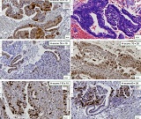
კეთილთვისებიანი/რეაქტიული დაზიანებები უპირატესად ავლენდა p16 შეღებვის ლაქოვან ან უარყოფით ნიმუშებს, ბუნებრივი ტიპის p53 ექსპრესიას და დაბალ პროლიფერაციულ აქტივობას, Ki67 ინდექსებით 5%-დან 6%-მდე. ამის საპირისპიროდ, ყველა სიმსივნისწინარე დაზიანება ავლენდა დიფუზურ ბლოკ-ტიპის p16 დადებითობას და p53 ჭარბ ექსპრესიას. Ki67 პროლიფერაციული აქტივობა მნიშვნელოვნად გაიზარდა სიმსივნისწინარე დაზიანებებში, 12%-დან 20%-მდე (p<0.001). პაციენტის საშუალო ასაკი უფრო მაღალი იყო სიმსივნისწინარე დაზიანებებში კეთილთვისებიან/რეაქტიულ დაზიანებებთან შედარებით. HPV დადებითობა უფრო ხშირად ასოცირდებოდა წინამორბედ დაზიანებებთან. p16, p53 და Ki67-ის კომბინირებული შეფასება ავლენდა მკაფიო იმუნოფენოტიპურ გამიჯვნას კეთილთვისებიან/რეაქტიულ და წინამორბედ ჯირკვლოვან დაზიანებებს შორის.
Downloads
Gogitidze, G.-J., Kepuladze, S., & Burkadze, G. (2025). Features of Neural Microenvironment Remodelling in cervical intraepithelial neoplasia and squamous Cell Carcinoma. Georgian Scientists, 7(2), 96–109. https://doi.org/10.52340/gs.2025.07.02.09
Gogitidze, G., Kepuladze, S., Tevzadze, N., & Burkadze, G. (2023). The Role of the Local Neural microenvironment in the Progression of cervical intraepithelial Neoplasia. Georgian Scientists, 5(3), 182–188. https://doi.org/10.52340/2023.05.03.19
Gogitidze, G.-J., Kepuladze, S., & Burkadze, G. (2025). Vascular Remodeling Across the Spectrum of Cervical Intraepithelial Neoplasia and Squamous Cell Carcinoma: A Digital Pathology-Based Morphometric and Immunohistochemical Analysis. Georgian Scientists, 7(2), 56–68. https://doi.org/10.52340/gs.2025.07.02.06
Adamashvili, N., Beriashvili, R., Tevzadze, N., Kepuladze, S., & Burkadze, G. (2024). Features of the distribution of acute and chronic inflammatory index in cervical intraepithelial neoplasia of different degrees and its relationship with proliferative activity detected by AgNOR technology. Georgian Scientists, 6(1), 45–57. https://doi.org/10.52340/gs.2024.06.01.08
Pirog EC, Kleter B, Olgac S, et al. Prevalence of human papillomavirus DNA in different histological subtypes of cervical adenocarcinoma. American Journal of Pathology 2000;157(4):1055–1062.
Stolnicu S, Talia KL, McCluggage WG. The Evolving Spectrum of Precursor Lesions of Cervical Adenocarcinomas. Adv Anat Pathol 2020;27(5):278–293.
Makino H, Sato S, Yajima A, Komatsu S, Fukao A. Evaluation of the effectiveness of cervical cancer screening: a case-control study in Miyagi, Japan. Tohoku J Exp Med 1995;175(3):171–178.
Boria F, Siegrist J, Hardisson D, Saturio N, Zapardiel I. Lobular endocervical glandular hyperplasia mimicking cervical adenocarcinoma. J Obstet Gynaecol (Lahore) 2021;41(7):1166–1168.
Bosch FX, Burchell AN, Schiffman M, et al. Epidemiology and Natural History of Human Papillomavirus Infections and Type-Specific Implications in Cervical Neoplasia. Vaccine 2008;26(SUPPL. 10).
Nara M, Hashi A, Murata SI, et al. Lobular endocervical glandular hyperplasia as a presumed precursor of cervical adenocarcinoma independent of human papillomavirus infection. Gynecol Oncol 2007;106(2):289–298.
Liao, S. Y., Rodgers, W. H., Kauderer, J., Darcy, K. M., Carter, R., Susumu, N., Nagao, S., Walker, J. L., Hatae, M., & Stanbridge, E. J. (2013). Endocervical glandular neoplasia associated with lobular endocervical glandular hyperplasia is HPV-independent and correlates with carbonic anhydrase-IX expression: a Gynaecological Oncology Group Study. British journal of cancer, 108(3), 613–620. https://doi.org/10.1038/bjc.2012.578
Endocervical glandular neoplasia associated with lobular endocervical glandular hyperplasia is HPV-independent and correlates with carbonic anhydrase-IX expression: a Gynaecological Oncology Group Study - PMC [Homepage on the Internet]. [cited 2026 May 19];Available from: https://pmc.ncbi.nlm.nih.gov/articles/PMC3593541/
Yamazaki H, Sasagawa T, Basha W, Segawa T, Inoue M. Hybrid capture-II and LCR-E7 PCR assays for HPV typing in cervical cytologic samples. Int J Cancer 2001;94(2):222–227.
Liao SY, Rodgers WH, Kauderer J, et al. Carbonic anhydrase IX (CA-IX) and high-risk human papillomavirus (H-HPV) as diagnostic biomarkers of cervical dysplasia/neoplasia in Japanese women with a cytologic diagnosis of atypical glandular cells (AGC): A Gynecologic Oncology Group (GOG) Study. Br J Cancer 2011;104(2):353–360.
Klaes R, Friedrich T, Spitkovsky D, et al. Overexpression of p16ink4a as a specific marker for dysplastic and neoplastic epithelial cells of the cervix uteri. Int J Cancer 2001;92(2):276–284.
Negri G, Egarter-Vigl E, Kasal A, Romano F, Haitel A, Mian C. P16INK4a is a useful marker for the diagnosis of adenocarcinoma of the cervix uteri and its precursors: An immunohistochemical study with immunocytochemical correlations. American Journal of Surgical Pathology 2003;27(2):187–193.
Mehdi HK, Raju K, Sheela SR. Association of P16, Ki-67, and CD44 expression in high-grade squamous intraepithelial neoplasia and squamous cell carcinoma of the cervix. J Cancer Res Ther 2023;19(8):S260–S267.
Benevolo M, Mottolese M, Marandino F, et al. Immunohistochemical expression of p16INK4a is predictive of HR-HPV infection in cervical low-grade lesions. Modern Pathology 2006;19(3):384–391.
Nucci MR. Pseudoneoplastic glandular lesions of the uterine cervix: A selective review. International Journal of Gynecological Pathology 2014;33(4):330–338.
Gilks CB, Young RH, Aguirre P, DeLellis RA, Scully RE. Adenoma malignum (minimal deviation adenocarcinoma) of the uterine cervix. A clinicopathological and immunohistochemical analysis of 26 cases. American Journal of Surgical Pathology 1989;13(9):717–729.
M Fischer MQLSKE. The p53-p21-DREAM-CDE/CHR pathway regulates G2/M cell cycle genes. Nucleic Acids Res 2016;44:164–174.
Blagih J, Buck MD, Vousden KH. p53, cancer and the immune response. J Cell Sci 2020;133(5).
საავტორო უფლებები (c) 2026 ქართველი მეცნიერები

ეს ნამუშევარი ლიცენზირებულია Creative Commons Attribution-NonCommercial-NoDerivatives 4.0 საერთაშორისო ლიცენზიით .