ეპითელურ-მეზენქიმული ტრანსფორმაციის მარკერების ექსპრესიის თავისებურებები საშვილოსნოს ყელის უმწიფარი პოლიპოიდური ბრტყელუჯრედოვანი მეტაპლაზის პროგრესირებაში


ჩამოტვირთვები


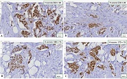
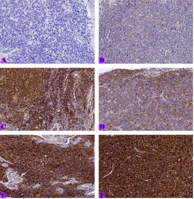
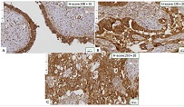
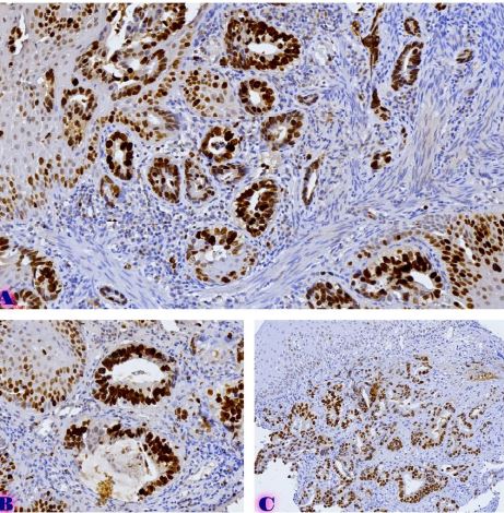
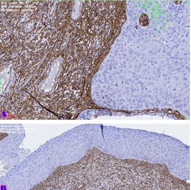
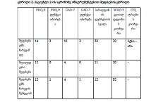
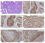
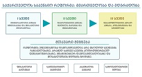
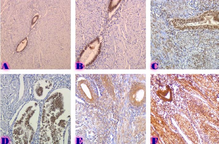
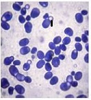
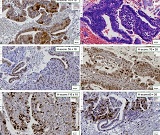
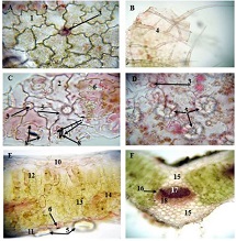
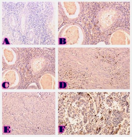
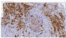
საშვილოსნოს ყელის ინტრაეპითელური ნეოპლაზია (CIN) არის საშვილოსნოს ყელის კიბოს მთავარი წინამორბედი და მისი პროგრესირების მოლეკულური მექანიზმების გაგება გადამწყვეტია დიაგნოსტიკური და თერაპიული სტრატეგიების გასაუმჯობესებლად. უმწიფარი პოლიპოიდური ბრტყელუჯრედოვანი მეტაპლაზია (IPSM) წარმოადგენს განსხვავებულ ჰისტოლოგიურ ერთეულს, რომელიც შესაძლოა პროგრესირებდეს CIN დაზიანებაში. ჩვენი კვლევა მიზნად ისახავდა ეპითელურ-მეზენქიმული ტრანსფორმაციის (EMT) მარკერების (ვიმენტინი, ბეტა-კატენინი და E-კადჰერინი) და ჰორმონალური რეცეპტორების (ესტროგენის რეცეპტორები [ER] და პროგესტერონის რეცეპტორების [PR]) ექსპრესიის შესწავლას IPSM-ისა და CIN-ის სხვადასხვა სტადიაში. ჩვენს კველაში ჩართული იყო სულ 195 პაციენტი მწიფე ბრტყელუჯრედოვანი მეტაპლაზიით, IPSM და CIN (CIN 1 და CIN 2). ჩატარდა იმუნოჰისტოქიმიური კვლევა ვიმენტინის, ბეტა-კატენინის, E-კადჰერინის, ER და PR-ის ექსპრესიის შესაფასებლად. ასევე ნაწილ პაციენტებში კლინიკურად განსაზღვრული იყო HPV სტატუსი და გაანალიზდა კორელაცია მარკერის ექსპრესიასა და დაზიანების ჰისტოლოგიურ ხარისხს შორის. ჩვენმა კვლევამ აჩვენა HPV-დადებით შემთხვევებში HPV–უარყოფით შემთხვევებთან შედარებით EMT მარკერების უფრო მაღალი ხარისხის ექსპრესია და E-cadherin-ის უფრო გამოხატული დაკარგვა. გარდა ამისა, ER და PR ექსპრესია მნიშვნელოვნად შემცირდა მაღალი ხარისხის CIN დაზიანებებში. ჩვენი კვლევის შედეგების საფუძველზე შეგვიძლია ვივარაუდოთ, რომ EMT მარკერების ექსპრესიის მომატება და ჰორმონალური რეცეპტორების დაქვეითება გარკვეულ როლს თამაშობს CIN-ისა და IPSM პროგრესირებაში. ეს მარკერები, განსაკუთრებით ვიმენტინი, ბეტა-კატენინი და E-კადჰერინი, შეიძლება იყოს ღირებული ბიომარკერი პროგრესირების რისკის სტრატიფიკაციისთვის საშვილოსნოს ყელის კიბოს პროფილაქტიკაში. დამატებითი კვლევებია საჭირო განსაკუთრებით HPV-დადებითი CIN დაზიანებების დროს ამ ბიომარკერების დამიზნებითი თერაპიული საშუალებების შემუშავებისათვის.
Downloads
R. Cappellesso et al., “The prognostic role of the epithelial-mesenchymal transition markers E-cadherin and Slug in laryngeal squamous cell carcinoma,” Histopathology, vol. 67, no. 4, pp. 491–500, Oct. 2015, doi: 10.1111/HIS.12668.
A. Auluck, G. Hislop, C. Bajdik, C. Poh, L. Zhang, and M. Rosin, “Trends in oropharyngeal and oral cavity cancer incidence of Human Papillomavirus (HPV)-related and HPV-unrelated sites in a multicultural population: The British Columbia experience,” Cancer, vol. 116, no. 11, pp. 2635–2644, Jun. 2010, doi: 10.1002/CNCR.25087.
N. A. Andrews, A. S. Jones, T. R. Helliwell, and A. R. Kinsella, “Expression of the E-cadherin-catenin cell adhesion complex in primary squamous cell carcinomas of the head and neck and their nodal metastases,” Br J Cancer, vol. 75, no. 10, pp. 1474–1480, 1997, doi: 10.1038/BJC.1997.252.
M. Lefevre et al., “Epithelial to mesenchymal transition and HPV infection in squamous cell oropharyngeal carcinomas: the papillophar study,” Br J Cancer, vol. 116, no. 3, p. 362, Jan. 2017, doi: 10.1038/BJC.2016.434.
S. Pirouzpanah, F. A. Taleban, P. Mehdipour, S. Sabour, and M. Atri, “Hypermethylation pattern of ESR and PgR genes and lacking estrogen and progesterone receptors in human breast cancer tumors: ER/PR subtypes,” Cancer Biomarkers, vol. 21, no. 3, pp. 621–638, 2018, doi: 10.3233/CBM-170697.
W. Truin, R. M. H. Roumen, S. Siesling, K. K. van de Vijver, V. C. G. Tjan-Heijnen, and A. C. Voogd, “Estrogen and progesterone receptor expression levels do not differ between lobular and ductal carcinoma in patients with hormone receptor-positive tumors,” Breast Cancer Res Treat, vol. 164, no. 1, pp. 133–138, Jul. 2017, doi: 10.1007/s10549-017-4220-x.
R. G. Lapidus, S. J. Nass, and N. E. Davidson, “The loss of estrogen and progesterone receptor gene expression in human breast cancer,” J Mammary Gland Biol Neoplasia, vol. 3, no. 1, pp. 85–94, 1998, doi: 10.1023/A:1018778403001.
M. Branca et al., “Upregulation of nuclear factor-κB (NF-κB) is related to the grade of cervical intraepithelial neoplasia, but is not an independent predictor of high-risk human papillomavirus or disease outcome in cervical cancer,” Diagn Cytopathol, vol. 34, no. 8, pp. 555–563, Aug. 2006, doi: 10.1002/dc.20514.
K. J. Syrjänen, “Spontaneous evolution of intraepithelial lesions according to the grade and type of the implicated human papillomavirus (HPV),” European Journal of Obstetrics and Gynecology and Reproductive Biology, vol. 65, no. 1, pp. 45–53, 1996, doi: 10.1016/0028-2243(95)02303-A.
M. J. Arends, C. H. Buckley, and M. Wells, “Aetiology, pathogenesis, and pathology of cervical neoplasia,” J Clin Pathol, vol. 51, no. 2, pp. 96–103, 1998, doi: 10.1136/jcp.51.2.96.
J. A. Glenn et al., “Longitudinal patterns of recurrence in patients with adrenocortical carcinoma,” Surgery (United States), vol. 165, no. 1, pp. 186–195, Jan. 2019, doi: 10.1016/J.SURG.2018.04.068.
M. Branca et al., “Up-regulation of proliferating cell nuclear antigen (PCNA) is closely associated with high-risk human papillomavirus (HPV) and progression of cervical intraepithelial neoplasia (CIN), but does not predict disease outcome in cervical cancer,” European Journal of Obstetrics and Gynecology and Reproductive Biology, vol. 130, no. 2, pp. 223–231, 2007, doi: 10.1016/j.ejogrb.2006.10.007.
M. Shen and Y. Kang, “Role Reversal: A Pro-metastatic Function of E-Cadherin,” Dev Cell, vol. 51, no. 4, pp. 417–419, Nov. 2019, doi: 10.1016/J.DEVCEL.2019.10.028.
T. Svanadze, S. Kepuladze, N. Tevzadze, and G. Burkadze, “ASSESSMENT OF PROLIFERATIVE ACTIVITY OF DIFFERENT TYPES OF SQUAMOUS CELL METAPLASIA OF THE CERVIX USING AgNor TECHNOLOGY,” ქართველი მეცნიერები, vol. 5, no. 2, pp. 275–287, Jun. 2023, doi: 10.52340/gs.2023.05.02.35.
S. Boulenouar et al., “Effects of HPV-16 E5, E6 and E7 proteins on survival, adhesion, migration and invasion of trophoblastic cells,” Carcinogenesis, vol. 31, no. 3, pp. 473–480, Mar. 2010, doi: 10.1093/CARCIN/BGP281.
საავტორო უფლებები (c) 2025 ქართველი მეცნიერები

ეს ნამუშევარი ლიცენზირებულია Creative Commons Attribution-NonCommercial-NoDerivatives 4.0 საერთაშორისო ლიცენზიით .