
MT1 Melatonin Receptor Expression Across Histological Types and Grades of Dysplasia in Colorectal Polyps

Downloads




Melatonin receptor-mediated signaling is involved in the regulation of cellular proliferation, apoptosis, and other processes potentially relevant to colorectal carcinogenesis. However, MT1 receptor expression across histological categories and grades of dysplasia in colorectal precursor lesions remains insufficiently characterized.
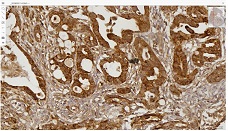
To evaluate immunohistochemical expression of the MT1 melatonin receptor in different histological types of colorectal polyps and determine its association with the presence and grade of dysplasia.
The study included 29 colorectal polyp specimens. Based on histological assessment, lesions were categorized as non-neoplastic polyps, serrated lesions, or conventional adenomas. Dysplasia was classified as absent, low-grade, or high-grade. MT1 immunohistochemical expression was quantified using a semiquantitative H-score (0–300), incorporating both the percentage of positive lesional epithelial cells and staining intensity. Between-group comparisons were performed using the Mann–Whitney U and Kruskal–Wallis tests. The association between increasing dysplasia grade and MT1 expression was assessed using Spearman rank correlation.
The median MT1 H-score in the overall cohort was 140 (IQR, 60–150). MT1 expression was significantly lower in dysplastic than in non-dysplastic lesions, with median H-scores of 40 (IQR, 40–45) and 140 (IQR, 120–160), respectively (p = 0.00020). MT1 expression also differed significantly across dysplasia grades (p = 0.000885). Increasing dysplasia grade showed a strong inverse correlation with MT1 H-score (Spearman ρ = −0.706; p = 0.000019). In contrast, MT1 expression did not differ significantly among the broad histological categories of non-neoplastic polyps, serrated lesions, and conventional adenomas (p = 0.383). No significant associations were identified between MT1 H-score and patient age, sex, or polyp size.
Reduced MT1 expression was associated with the presence and increasing grade of dysplasia in colorectal polyps, whereas no independent difference was detected among broad histological categories. These findings indicate that altered MT1 expression may be more closely associated with the dysplastic phenotype than with histological category alone. Given the limited cohort size, the potential role of MT1 in early colorectal neoplastic progression requires evaluation in larger cohorts with more detailed histological stratification.
Downloads
[1] R. L. Siegel et al., “Colorectal cancer statistics, 2020,” CA Cancer J. Clin., vol. 70, no. 3, pp. 145–164, May 2020, doi: 10.3322/CAAC.21601.
[2] N. Arber et al., “Celecoxib for the prevention of colorectal adenomatous polyps,” New Engl. J. Med., vol. 355, no. 9, pp. 885–895, Aug. 2006, doi: 10.1056/nejmoa061652.
[3] F. M. Giardiello et al., “Primary chemoprevention of familial adenomatous polyposis with sulindac,” New Engl. J. Med., vol. 346, no. 14, pp. 1054–1059, Apr. 2002, doi: 10.1056/nejmoa012015.
[4] H. Ishiguro et al., “NOTCH1 activates the Wnt/β-catenin signaling pathway in colon cancer,” Oncotarget, vol. 8, no. 36, pp. 60378–60389, 2017, doi: 10.18632/ONCOTARGET.19534.
[5] M. L. Dubocovich and M. Markowska, “Functional MT1 and MT2 melatonin receptors in mammals,” Endocrine, vol. 27, no. 2, pp. 101–110, 2005, doi: 10.1385/ENDO:27:2:101.
[6] P. T. Ram et al., “Involvement of the mt1 melatonin receptor in human breast cancer,” Cancer Lett., vol. 179, no. 2, pp. 141–150, May 2002, doi: 10.1016/S0304-3835(01)00873-4.
[7] J. Liu, S. J. Clough, A. J. Hutchinson, E. B. Adamah-Biassi, M. Popovska-Gorevski, and M. L. Dubocovich, “MT1 and MT2 Melatonin Receptors: A Therapeutic Perspective,” Annu. Rev. Pharmacol. Toxicol., vol. 56, p. 361, Jan. 2015, doi: 10.1146/ANNUREV-PHARMTOX-010814-124742.
[8] J. León et al., “Gender-related invasion differences associated with mRNA expression levels of melatonin membrane receptors in colorectal cancer,” Mol. Carcinog., vol. 51, no. 8, pp. 608–618, Aug. 2012, doi: 10.1002/mc.20832.
[9] K. Jablonska et al., “Expression of the MT1 Melatonin Receptor in Ovarian Cancer Cells,” Int. J. Mol. Sci., vol. 15, no. 12, p. 23074, Dec. 2014, doi: 10.3390/IJMS151223074.
[10] T. B. de Castro, A. L. Mota, N. A. Bordin-Junior, D. S. Neto, and D. A. P. C. Zuccari, “Immunohistochemical Expression of Melatonin Receptor MT1 and Glucose Transporter GLUT1 in Human Breast Cancer,” Anticancer Agents Med. Chem., vol. 18, no. 15, pp. 2110–2116, Feb. 2019, doi: 10.2174/1871520618666181025125532.
[11] M. Chen et al., “Melatonin MT1 and MT2 receptor ERK signaling is differentially dependent on Gi/o and Gq/11 proteins,” J. Pineal Res., vol. 68, no. 4, May 2020, doi: 10.1111/JPI.12641.
[12] Y. Lecarpentier, O. Schussler, J. L. Hébert, and A. Vallée, “Multiple Targets of the Canonical WNT/β-Catenin Signaling in Cancers,” Front. Oncol., vol. 9, Nov. 2019, doi: 10.3389/FONC.2019.01248.
[13] A. Debebe et al., “Wnt/β-catenin activation and macrophage induction during liver cancer development following steatosis,” Oncogene, vol. 36, no. 43, pp. 6020–6029, Oct. 2017, doi: 10.1038/onc.2017.207.
[14] A. Ring, Y. M. Kim, and M. Kahn, “Wnt/Catenin Signaling in Adult Stem Cell Physiology and Disease,” Stem Cell Rev. Rep., vol. 10, no. 4, pp. 512–525, Aug. 2014, doi: 10.1007/S12015-014-9515-2.
Copyright (c) 2026 Georgian Scientists

This work is licensed under a Creative Commons Attribution-NonCommercial-NoDerivatives 4.0 International License.