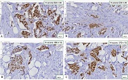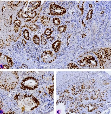
ASSESSMENT OF PROLIFERATIVE ACTIVITY OF IMMATURE OVARIAN TERATOMAS USING AgNOR TECHNOLOGY

Downloads


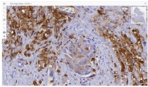
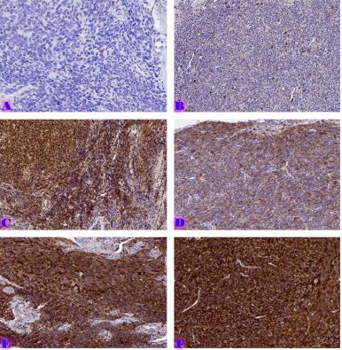
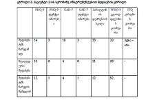
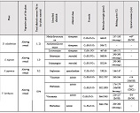
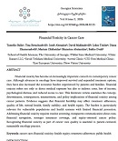
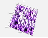
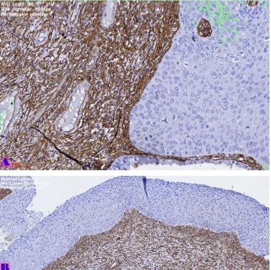
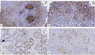
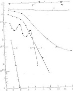
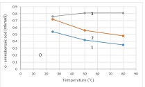
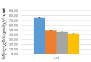
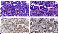
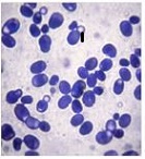
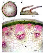
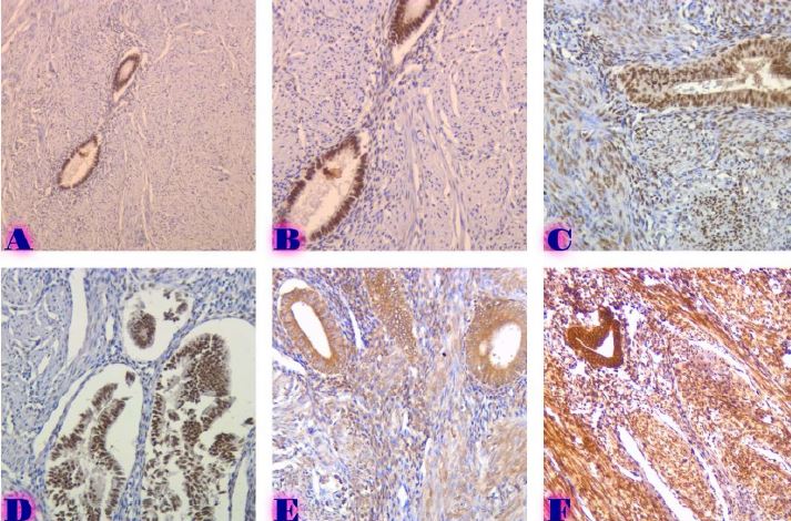
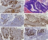
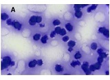
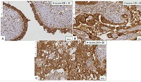
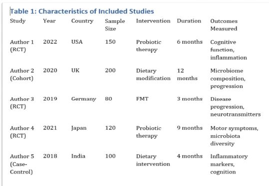
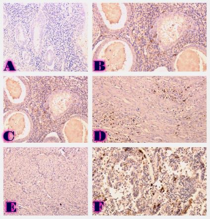
Germ cell tumors arise from the covering stem cells on the surface of the embryonic ovary. They make up 20-30% of all types of ovarian tumors. About 95% are benign and present as mature cystic teratoma, and only 5% are malignant. Malignant germ cell tumors of the ovary represent 2.6% of all malignant tumors of the ovary, in contrast to epithelial tumors of the ovary (95%). A high incidence of ovarian malignant germ cell tumors is observed in the first two decades of life. Determining the prognosis of germ cell tumors of the ovary is a complex and problematic issue, and according to the existing literature, various methods are used to determine a more accurate classification of cases. One of them can be used AgNOR staining, which is called one of the means of proliferation assessment in the case of different tumors, as well as in the differentiation of dysplasias and benign and malignant processes. According to our research, it is used to evaluate proliferative activity during different histological differentiation of immature teratomas.
Downloads
Ulbright TM. Germ cell tumors of the gonads: A selective review emphasizing problems in differential diagnosis, newly appreciated, and controversial issues. Modern Pathology. 2005 Feb;18(SUPPL. 2).
Jorge S, Jones NL, Chen L, Hou JY, Tergas AI, Burke WM, et al. Characteristics, Treatment and Outcomes of Women with Immature Ovarian Teratoma, 1998–2012. Gynecol Oncol [Internet]. 2016 Aug 1 [cited 2023 Mar 20];142(2):261. Available from: /pmc/articles/PMC4961548/
Outwater EK, Siegelman ES, Hunt JL. Ovarian Teratomas: Tumor Types and Imaging Characteristics. Radiographics. 2001;21(2):475–90.
Javadi S, Ganeshan DM, Qayyum A, Iyer RB, Bhosale P. Ovarian cancer, the revised FIGO staging system, and the role of imaging. American Journal of Roentgenology. 2016 Jun 1;206(6):1351–60.
BLACKWELL WJ, DOCKERTY MB. Dermoid cysts of the ovary: their clinical and pathologic significance. Am J Obstet Gynecol. 1946 Feb 1;51(2):151–72.
Meliti A, Hafiz B, Al-Maghrabi H, Gari A. Collision Glial Neoplasms Arising in an Ovarian Mature Cystic Teratoma: A Rare Event. Case Rep Pathol. 2020 Feb 3;2020:1–4.
Nogales FF, Dulcey I, Preda O. Germ cell tumors of the ovary: An update. Arch Pathol Lab Med. 2014;138(3):351–62.
Fellegara G, Young RH, Kuhn E, Rosai J. Ovarian mature cystic teratoma with florid vascular proliferation and Wagner-Meissner-like corpuscles. Int J Surg Pathol. 2008 Jul;16(3):320–3.
Murdock T, Orr B, Allen S, Ibrahim J, Sharma R, Ronnett BM, et al. Central Nervous System-type Neuroepithelial Tumors and Tumor-like Proliferations Developing in the Gynecologic Tract and Pelvis. American Journal of Surgical Pathology. 2018 Nov 1;42(11):1429–44.
Tannapfel A, Nüßlein S, Fietkau R, Katalinic A, Köckerling F, Wittekind C. Apoptosis, proliferation, bax, bcl-2 and p53 status prior to and after preoperative radiochemotherapy for locally advanced rectal cancer. Int J Radiat Oncol Biol Phys. 1998 Jun 1;41(3):585–91.
Mele AA, Danak S, Ellis E, Green A, Mele AA, Danak SU, et al. Immature Teratoma With Metastatic Gliosis. Cureus [Internet]. 2022 Mar 1 [cited 2023 Mar 20];14(3). Available from: https://www.cureus.com/articles/85769-immature-teratoma-with-metastatic-gliosis
Gheorghisan-Galateanu A, Terzea DC, Carsote M, Poiana C. Immature ovarian teratoma with unusual gliomatosis. J Ovarian Res. 2013;6(1).
Ferreira SJ, Machado MÂN, de Lima AAS, Johann ACBR, Grégio AMT, Azevedo-Alanis LR. Identification of AgNORs and cytopathological changes in oral lichen planus lesions. Acta Histochem. 2017 Jan 1;119(1):32–8.
Harsh A, Tondon R, Harsh HK, Professor E. Utility of AgNOR Count in Non-Neoplastic and Neoplastic Lesions of the Uterine Cervix. Original Research Article [Internet]. 2018 [cited 2022 Dec 30];198(1):198–201. Available from: www.ijmrp.com
Aggarwal T, Sawke G, Sawke N. Application of AgNOR (Argyrophilic Nucleolar Organizer Regions) Staining in Distinction of Non Neoplastic and Neoplastic Endometrial Lesions INTRODUCTION. People’s Journal of Scientific Research. 2015;8(1).

This work is licensed under a Creative Commons Attribution-NonCommercial-NoDerivatives 4.0 International License.