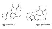
Features of Dentin-Enamel junction in different types of teeth and designated age groups

Downloads


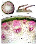
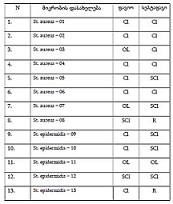
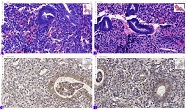
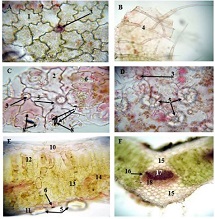
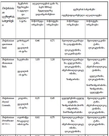
Dentin-Enamel Junction is an anatomical interfacial region between the dentin and outer enamel coating in teeth. The uniqueness of this region is due to the incorporation of three morphologically different strong tissue of teeth. It has the utmost importance in clinical Dentistry as well as dental X-rays therefore needs to be cautiously addressed in routine dental practice. There are distinguished four types of connection within the cervical region of teeth: type 1 - cementum is partially covering enamel; Type 2 - Dentin-enamel is attached through endings "Margin-to-margin'' connection; Type 2 - cementum and enamel are not directly connected and there is denuded region up the teeth root. Type 4 -the enamel is covering the cementum; The main goal of our research was to study the different features of dentin-enamel Junction in various types of teeth and age groups. We studied 280 teeth which were assigned into four types of the group according to teeth types and three groups according to age: Group 1 - incisors (60 teeth); Group 2-canine (60 teeth); Group 3 - Premolars (80 teeth) in designated three age groups within the age intervals: 16-30 Y., 30-50Y and 50-70Y.
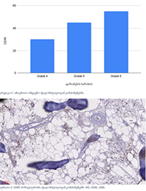
According to the results of our research, in all types of teeth dominant type of dental-enamel Junction is type 3. The total percentage is 43.8%, and the frequency is increasing along with the rise in age and the maximum is reached in 50-70 year intervals respectively. This type of junction is also the most common in canines and has an equal frequency in molars and premolars. In all types of teeth the dental-enamel junction subtype is in distributed : Type 1 - 22,9%, Type 2 - 30%, Type 3 - 42,8%, Type 4 - 4,3%. besides that, type 1 junction in the 1 and 2 research group (molars and canine) has the same frequency in all designated age groups. Type 4 junction was not detected in any cases in the whole group neither in Group 2 nor in 50-70 years of intervals.
Downloads
Zhang C, Mo D, Guo J, Wang W, Long S, Zhu H, Chen D, Ge G, Tang Y. A method of crack detection based on digital image correlation for simulated cracked tooth. BMC Oral Health. 2021 Oct 19;21(1):539. doi: 10.1186/s12903-021-01897-2. PMID: 34666731; PMCID: PMC8524926.
Bi R, Lyu P, Song Y, Li P, Song D, Cui C, Fan Y. Function of Dental Follicle Progenitor/Stem Cells and Their Potential in Regenerative Medicine: From Mechanisms to Applications. Biomolecules. 2021 Jul 7;11(7):997. doi: 10.3390/biom11070997. PMID: 34356621; PMCID: PMC8301812.
Nascimento MM, Dilbone DA, Pereira PN, Duarte WR, Geraldeli S, Delgado AJ. Abfraction lesions: etiology, diagnosis, and treatment options. Clin Cosmet Investig Dent. (2016 )May 3;8:79-87. doi: 10.2147/CCIDE.S63465. PMID: 27217799; PMCID: PMC4861607.
Bosshardt DD, Stadlinger B, Terheyden H. Cell-to-cell communication--periodontal regeneration. Clin Oral Implants Res. 2015 Mar;26(3):229-39. doi: 10.1111/clr.12543. Epub 2015 Jan 2. PMID: 25639287.
Zucchelli, Giovanni, Guido Gori, Monica Mele, Martina Stefanini, Claudio Mazzotti, Matteo Marzadori, Lucio Montebugnoli, and Massimo De Sanctis. "Non‐carious cervical lesions associated with gingival recessions: A decision‐making process." Journal of Periodontology 82, no. 12 (2011): 1713-1724.
Tatakis DN, Chambrone L, Allen EP, Langer B, McGuire MK, Richardson CR, Zabalegui I, Zadeh HH. Periodontal soft tissue root coverage procedures: a consensus report from the AAP Regeneration Workshop. J Periodontol. 2015 Feb;86(2 Suppl):S52-5. doi: 10.1902/jop.2015.140376. Epub 2014 Oct 15. PMID: 25315018.
Arambawatta, Kapila, Roshan Peiris, and Deepthi Nanayakkara. "Morphology of the cemento-enamel junction in premolar teeth." Journal of oral science 51.4 (2009): 623-627.
Ceppi, E., et al. "Cementoenamel junction of deciduous teeth: SEM-morphology." Eur J Paediatr Dent 7.3 (2006): 131-4.
Zeichner-David M, Oishi K, Su Z, Zakartchenko V, Chen LS, Arzate H, Bringas P Jr. Role of Hertwig's epithelial root sheath cells in tooth root development. Dev Dyn. 2003 Dec;228(4):651-63. doi: 10.1002/dvdy.10404. PMID: 14648842.
Neuvald, Lilian, and Alberto Consolaro. "Cementoenamel junction: microscopic analysis and external cervical resorption." Journal of Endodontics 26.9 (2000): 503-508. [11] Leonardi, R., Loreto, C., Caltabiano, R., & Caltabiano, C. (1996). The cervical third of deciduous teeth. An ultrastructural study of the heard tissues by SEM. Minerva Stomatologica, 45(3), 75-79.
Muller, C. J., and C. W. Van Wyk. "The amelo-cemental junction." The Journal of the Dental Association of South Africa= Die Tydskrif van die Tandheelkundige Vereniging van Suid-Afrika 39.12 (1984): 799-803.

This work is licensed under a Creative Commons Attribution-NonCommercial-NoDerivatives 4.0 International License.